Ventricles Of The Brain Functions Of The Ventricular 50 Off

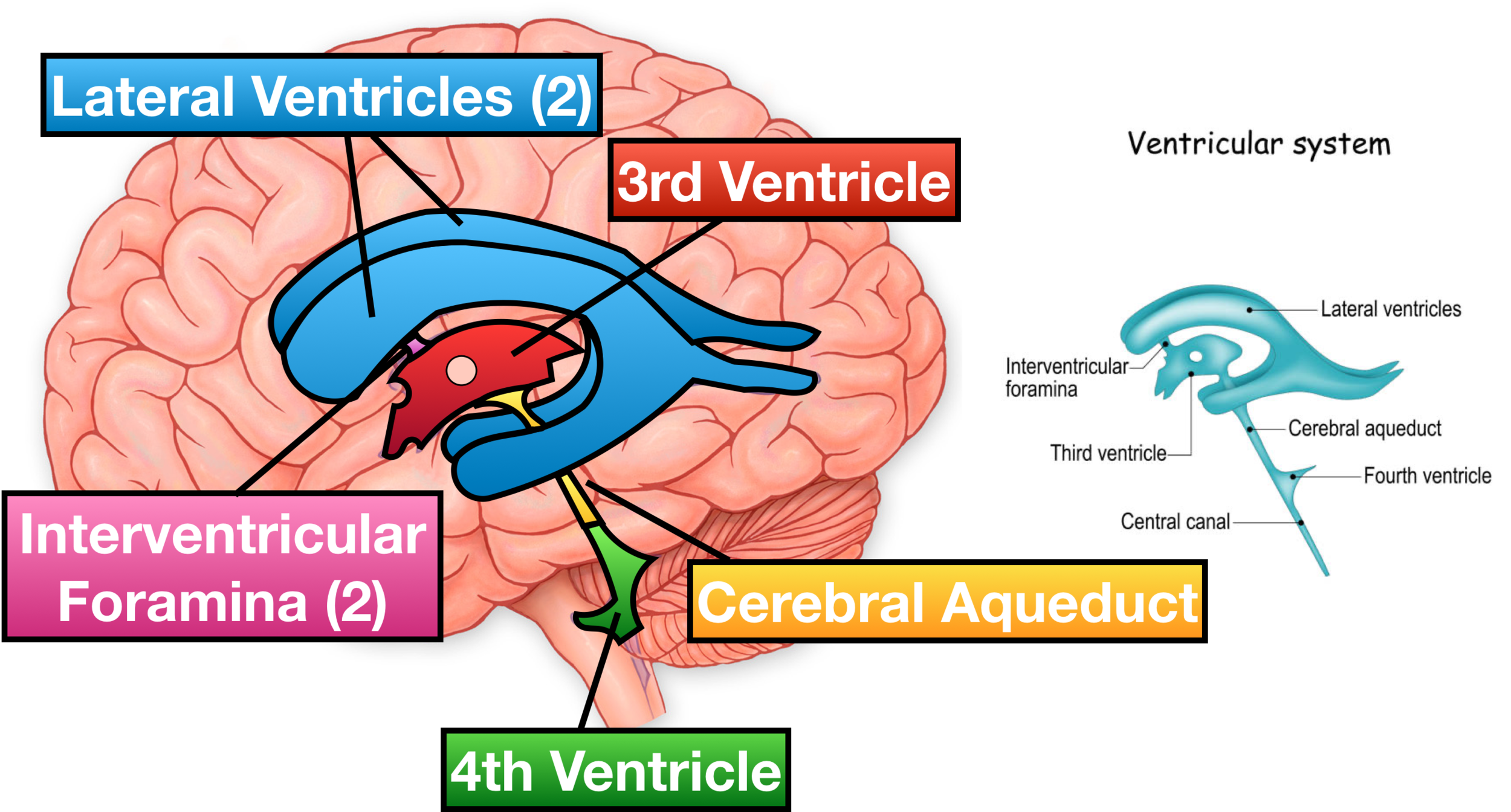
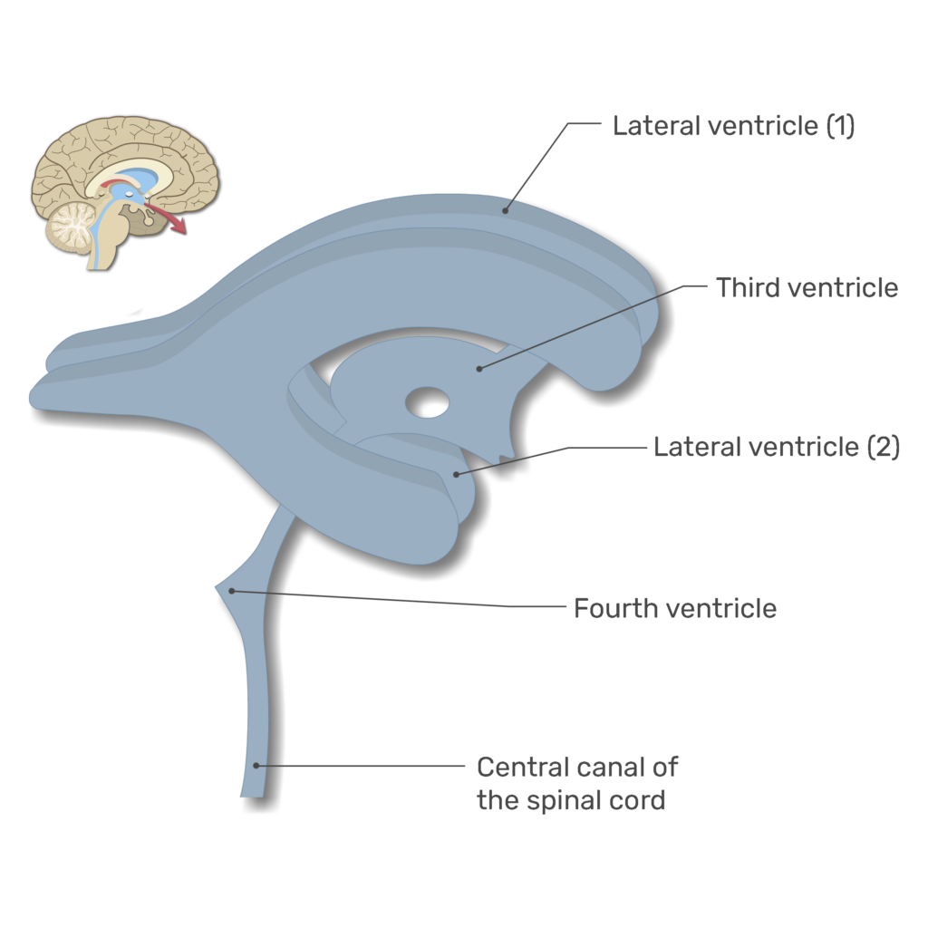
Ventricles Of The Brain Labeled Anatomy Function Csf Flow Aside from cerebrospinal fluid, your brain ventricles are hollow. their sole function is to produce and secrete cerebrospinal fluid to protect and maintain your central nervous system. csf is constantly bathing the brain and spinal column, clearing out toxins and waste products released by nerve cells. Ventricular system anatomy. the ventricular system is a network of communicating cavities within the brain, called ventricles, that function to produce, transport, and reabsorb cerebrospinal fluid (csf) throughout the central nervous system. the main structures that make up the ventricular system include:.

Ventricles Of The Brain Functions Of The Ventricular 50 Off Ventricles of the brain. ventricular system of the brain with neighboring structures. the human brain is so vital and delicate that it is fully encased in a bony vault in order to protect it from damage. to add even more protection, the brain is wrapped in three meningeal layers – dura mater, arachnoid mater and pia mater. The ventricular system consists of four ventricles in the brain. ventricles are a communicating network of chambers filled with cerebrospinal fluid (csf). the ventricular system is the pathway for the csf and is critical to the central nervous system ’s overall functioning. developmental anomalies that impact the ventricular system include. The lateral ventricles are connected to the third ventricle by the foramen of monro. the third ventricle is situated in between the right and the left thalamus. the anterior surface of the ventricle contains two protrusions: supra optic recess – located above the optic chiasm. infundibular recess – located above the optic stalk. The ventricular system of the brain is an interconnected series of cavities filled with cerebrospinal fluid (csf) that cushions the brain. though the presence of cerebral ventricles was known since ancient times, its function was obscure. early scientists believed ventricles to be the site of thought, emotions, reasoning, and memory. leonardo da vinci, the famed artist who drew mona lisa.

Ventricles Of The Brain Anatomy And Cerebrospinal Fluid Csf The lateral ventricles are connected to the third ventricle by the foramen of monro. the third ventricle is situated in between the right and the left thalamus. the anterior surface of the ventricle contains two protrusions: supra optic recess – located above the optic chiasm. infundibular recess – located above the optic stalk. The ventricular system of the brain is an interconnected series of cavities filled with cerebrospinal fluid (csf) that cushions the brain. though the presence of cerebral ventricles was known since ancient times, its function was obscure. early scientists believed ventricles to be the site of thought, emotions, reasoning, and memory. leonardo da vinci, the famed artist who drew mona lisa. Overview. the ventricles of the brain are a communicating network of cavities filled with cerebrospinal fluid (csf) and located within the brain parenchyma. the ventricular system is composed of 2 lateral ventricles, the third ventricle, the cerebral aqueduct, and the fourth ventricle (see the images below). the choroid plexuses are located in. The cerebral ventricles are a series of interconnected, fluid filled spaces that lie in the core of the forebrain and brainstem (figure 1.17). the presence of ventricular spaces in the various subdivisions of the brain reflects the fact that the ventricles are the adult derivatives of the open space or lumen of the embryonic neural tube (see chapter 22).

Comments are closed.