Ventricle Model Labeled Google Search Nervous System Anatomy

Brain Ventricle Anatomy Diagram Image Radiopaedia Org The ventricles are structures that produce cerebrospinal fluid, and transport it around the cranial cavity. they are lined by ependymal cells, which form a structure called the choroid plexus. it is within the choroid plexus that csf is produced. embryologically, the ventricular system is derived from the lumen of the neural tube. This interactive brain model is powered by the wellcome trust and developed by matt wimsatt and jack simpson; reviewed by john morrison, patrick hof, and edward lein. structure descriptions were written by levi gadye and alexis wnuk and jane roskams .

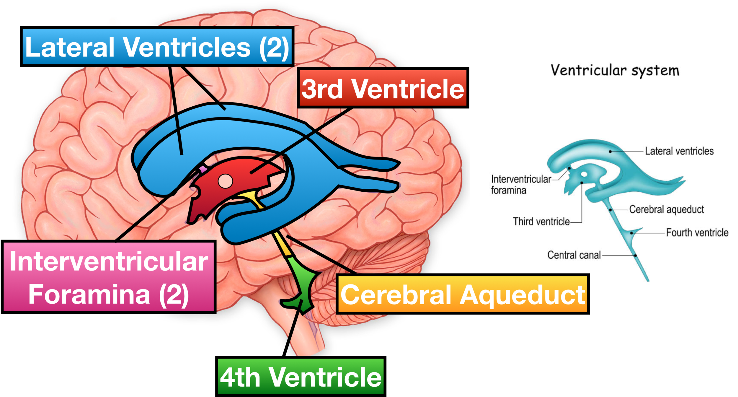
Ventricles Of The Brain Labeled Anatomy Function Csf Flow Aside from cerebrospinal fluid, your brain ventricles are hollow. their sole function is to produce and secrete cerebrospinal fluid to protect and maintain your central nervous system. csf is constantly bathing the brain and spinal column, clearing out toxins and waste products released by nerve cells. Ventricles of the brain. ventricular system of the brain with neighboring structures. the human brain is so vital and delicate that it is fully encased in a bony vault in order to protect it from damage. to add even more protection, the brain is wrapped in three meningeal layers – dura mater, arachnoid mater and pia mater. The ventricles of the brain are the four cavities containing cerebrospinal fluid (csf). namely, they are: two lateral ventricles, often called the first and second ventricles; third ventricle; fourth ventricle; the ventricles are connected to each other and to the central canal of the spinal cord, comprising the ventricular system of the brain. Quick facts. the ventricular system of the brain consists of four ventricular chambers, which permeate the brain. these include the right and left lateral ventricles, (also known as the first and second ventricles), the third ventricle and the fourth ventricle. they contain the cerebrospinal fluid (csf) which cushions the brain.

Ventricle Model Labeled Google Search Nervous System Anatomy The ventricles of the brain are the four cavities containing cerebrospinal fluid (csf). namely, they are: two lateral ventricles, often called the first and second ventricles; third ventricle; fourth ventricle; the ventricles are connected to each other and to the central canal of the spinal cord, comprising the ventricular system of the brain. Quick facts. the ventricular system of the brain consists of four ventricular chambers, which permeate the brain. these include the right and left lateral ventricles, (also known as the first and second ventricles), the third ventricle and the fourth ventricle. they contain the cerebrospinal fluid (csf) which cushions the brain. Ventricular system anatomy. the ventricular system is a network of communicating cavities within the brain, called ventricles, that function to produce, transport, and reabsorb cerebrospinal fluid (csf) throughout the central nervous system. the main structures that make up the ventricular system include: lateral ventricles (2). 242787. anatomical terms of neuroanatomy. [ edit on wikidata] in neuroanatomy, the ventricular system is a set of four interconnected cavities known as cerebral ventricles in the brain. [ 1 ][ 2 ] within each ventricle is a region of choroid plexus which produces the circulating cerebrospinal fluid (csf).

Human Brain Anatomy And Function Cerebrum Brainstem Ventricular system anatomy. the ventricular system is a network of communicating cavities within the brain, called ventricles, that function to produce, transport, and reabsorb cerebrospinal fluid (csf) throughout the central nervous system. the main structures that make up the ventricular system include: lateral ventricles (2). 242787. anatomical terms of neuroanatomy. [ edit on wikidata] in neuroanatomy, the ventricular system is a set of four interconnected cavities known as cerebral ventricles in the brain. [ 1 ][ 2 ] within each ventricle is a region of choroid plexus which produces the circulating cerebrospinal fluid (csf).

Comments are closed.