Ventricle Model For Brain Series

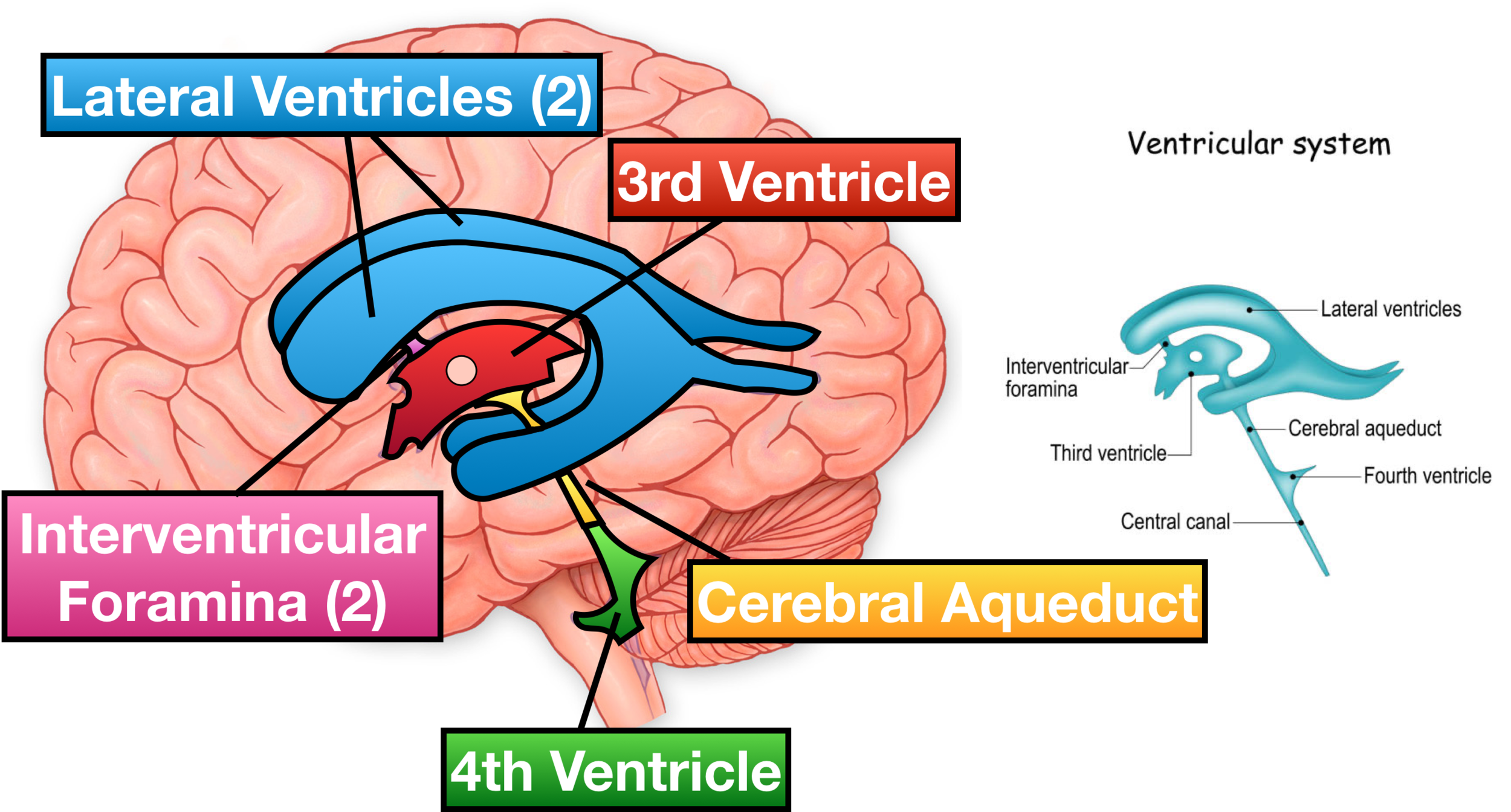
Ventricles Of The Brain Labeled Anatomy Function Csf Flow This video provides a brief overview of the ventricle model.structures included:lateral ventricleinter ventricular foramenthird ventriclecerebral aqueduct4th. This interactive brain model is powered by the wellcome trust and developed by matt wimsatt and jack simpson; reviewed by john morrison, patrick hof, and edward lein. structure descriptions were written by levi gadye and alexis wnuk and jane roskams .

Ventricle Model For Brain Series Youtube 05 the ventricles (27 minutes) the ventricles are demonstrated and named on a model cast as well as in rotating 3d reconstructions. the production, function, circulation and removal of csf produced by the choroid plexus is discussed using a diagram and then reviewed on frontal, axial and sagittal brain specimens and corresponding mri’s. 242787. anatomical terms of neuroanatomy. [ edit on wikidata] in neuroanatomy, the ventricular system is a set of four interconnected cavities known as cerebral ventricles in the brain. [ 1 ][ 2 ] within each ventricle is a region of choroid plexus which produces the circulating cerebrospinal fluid (csf). This page titled 11.6: models 3d brains, ventricles, sagittal and coronal brain slices, torsos, sagittal head is shared under a cc by nc sa 4.0 license and was authored, remixed, and or curated by laird c. sheldahl via source content that was edited to the style and standards of the libretexts platform. Updated on january 20, 2019. the ventricular system is a series of connecting hollow spaces called ventricles in the brain that are filled with cerebrospinal fluid. the ventricular system consists of two lateral ventricles, the third ventricle, and the fourth ventricle. the cerebral ventricles are connected by small pores called foramina, as.

Ventricles Of The Brain Anatomy And Cerebrospinal Fluid Csf This page titled 11.6: models 3d brains, ventricles, sagittal and coronal brain slices, torsos, sagittal head is shared under a cc by nc sa 4.0 license and was authored, remixed, and or curated by laird c. sheldahl via source content that was edited to the style and standards of the libretexts platform. Updated on january 20, 2019. the ventricular system is a series of connecting hollow spaces called ventricles in the brain that are filled with cerebrospinal fluid. the ventricular system consists of two lateral ventricles, the third ventricle, and the fourth ventricle. the cerebral ventricles are connected by small pores called foramina, as. The ventricular system of the brain is an interconnected series of cavities filled with cerebrospinal fluid (csf) that cushions the brain. though the presence of cerebral ventricles was known since ancient times, its function was obscure. early scientists believed ventricles to be the site of thought, emotions, reasoning, and memory. leonardo da vinci, the famed artist who drew mona lisa. Ventricles of the brain. ventricular system of the brain with neighboring structures. the human brain is so vital and delicate that it is fully encased in a bony vault in order to protect it from damage. to add even more protection, the brain is wrapped in three meningeal layers – dura mater, arachnoid mater and pia mater.

Comments are closed.