Ultra Widefield Imaging Numerous Retinal Pathologies Obn

Ultra Widefield Imaging Numerous Retinal Pathologies Obn Abstract. the development of ultra widefield retinal imaging has accelerated our understanding of common retinal diseases. as we continue to validate the diagnostic and prognostic significance of pathology in the retinal periphery, the ability to visualize and evaluate these features in an efficient and patient friendly manner will become more. Traditional colour fundus photography (cfp) typically images 30–60° of the retina, but new imaging devices allow for ultra widefield (uwf) retinal images with a field of view of 100–200.

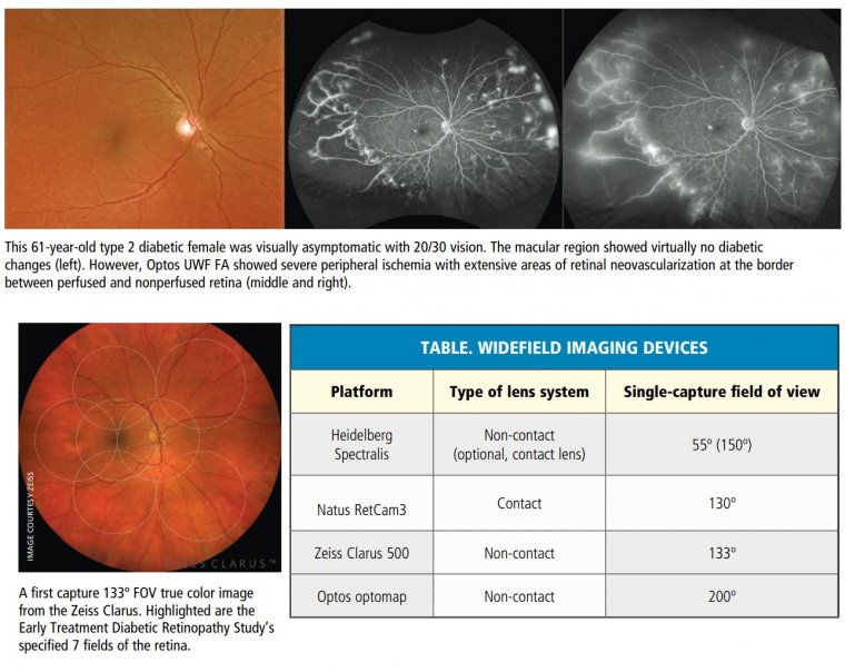
Ultra Widefield Retinal Imaging Noosa Optical Noosa Junction In 2019, the international widefield imaging study group defined widefield imaging as a field of view of approximately 60 to 100 degrees, capturing the mid periphery of the retina up to the posterior edge of the vortex vein ampulla. 1 it defined ultra widefield imaging as an image of the far periphery of the retina, including the anterior edge of the vortex vein ampulla and beyond. 1 this. Fundus photography in the modern era has consisted of imaging systems capable of visualizing between 30° to 45° of the retina. widefield imaging systems are defined as having the capability to visualize 50° or more, while the diabetic retinopathy clinical research network, a renowned research and imaging organization, defines ultra widefield. Uwf imaging devices produce high resolution images of the retina, including the far periphery beyond the vortex ampullae. uwf imaging was introduced about 30 years ago, after a 5 year old boy was blinded in one eye following a regular eye exam that failed to spot a retinal detachment. determined to protect others from similar experiences, the. While traditional fundus cameras are able to capture approximately 30 50° of the retina, ultra widefield fundus imaging is able to capture a wider field of up to 200° of the retina.

Ultra Widefield Fundus Photography Brisbane Eye Doctor Clinic Uwf imaging devices produce high resolution images of the retina, including the far periphery beyond the vortex ampullae. uwf imaging was introduced about 30 years ago, after a 5 year old boy was blinded in one eye following a regular eye exam that failed to spot a retinal detachment. determined to protect others from similar experiences, the. While traditional fundus cameras are able to capture approximately 30 50° of the retina, ultra widefield fundus imaging is able to capture a wider field of up to 200° of the retina. Figure 1. a traditional fundus photograph of subretinal hemorrhage in the macula. definition and devices. the diabetic retinopathy clinical research network (drcr network) initially defined uwf as having at least a 100° view of the retina. 4 the current consensus definition from the international widefield imaging study group states that wf imaging captures images of the retina posteriorly to. The development of ultra widefield retinal imaging has accelerated our understanding of common retinal diseases. as we continue to validate the diagnostic and prognostic significance of pathology in the retinal periphery, the ability to visualize and evaluate these features in an efficient and patient friendly manner will become more important.

Optos Ultra Widefield Retinal Imaging System Mivision Figure 1. a traditional fundus photograph of subretinal hemorrhage in the macula. definition and devices. the diabetic retinopathy clinical research network (drcr network) initially defined uwf as having at least a 100° view of the retina. 4 the current consensus definition from the international widefield imaging study group states that wf imaging captures images of the retina posteriorly to. The development of ultra widefield retinal imaging has accelerated our understanding of common retinal diseases. as we continue to validate the diagnostic and prognostic significance of pathology in the retinal periphery, the ability to visualize and evaluate these features in an efficient and patient friendly manner will become more important.

Comments are closed.