The Ovaries Structure Ligaments Vascular Supply Function

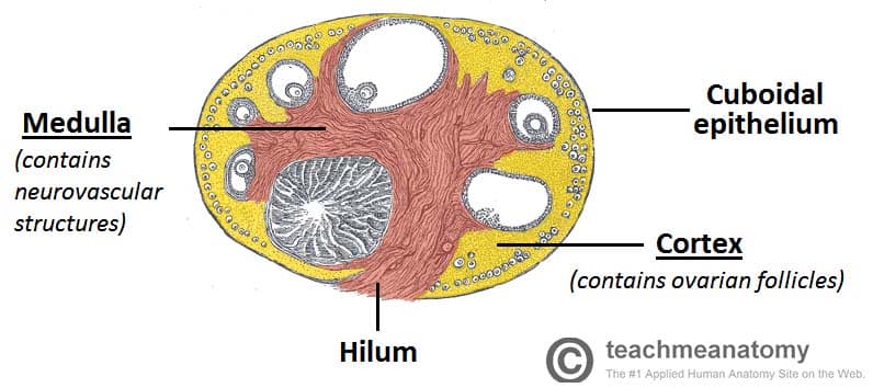
The Ovaries Structure Ligaments Vascular Supply Function The ovaries are paired, oval organs attached to the posterior surface of the broad ligament of the uterus by the mesovarium (a fold of peritoneum, continuous with the outer surface of the ovaries). neurovascular structures enter the hilum of the ovary via the mesovarium. the main functions of the ovaries are: to produce oocytes (female gametes. Structure morphology. the ovaries are approximately 4x2x3cm in size, whitish, and consist of an inner medulla and an outer cortex (standring, 2016). the cortex surrounds the medulla except at the hilum, where the ovarian vessels and nerves enter the ovary. the cortex itself is covered by a collagenous tunica albuginea and an epithelial layer.

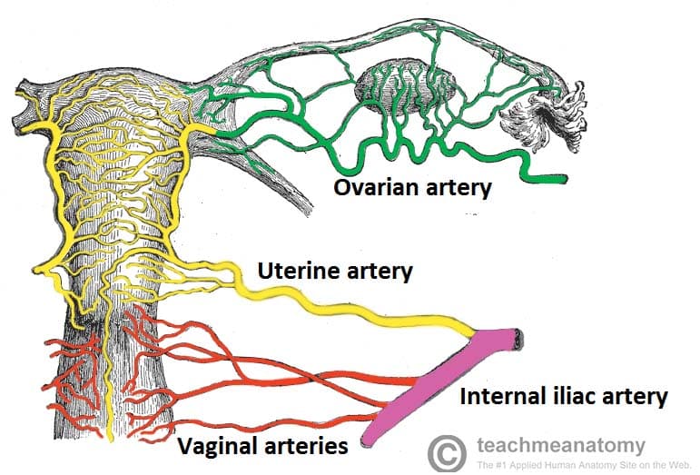
The Ovaries Structure Ligaments Vascular Supply Function The ligaments of the female reproductive tract are a series of structures that support the internal female genitalia in the pelvis. the ligaments of the female reproductive tract can be divided into three categories: broad ligament – a sheet of peritoneum, associated with both the uterus and ovaries. uterine ligaments – ligaments primarily. The ovarian artery is a paired structure that originates from the abdominal aorta below the renal artery or around l2. the artery runs through the suspensory ligament of the ovary, which then enters the mesovarium. the ovarian artery may make an anastomosis with the uterine artery within the broad ligament. The suspensory ligament of the ovary, also known as the infundibulopelvic ligament, is a thin fold of parietal peritoneum extending from the lateral margin of the ovary and to the lateral wall of the female pelvis. it makes up the superior border of the broad ligament of the uterus and helps to anchor the ovary to the posterior pelvic wall. The ligament of the ovary progresses as the round ligament (both of which are derived from the gubernaculum), which secures each ovary to the cornu of the uterus. furthermore, each round ligament also attaches to the inner part of the labia majora. the mesovarium, which is a double layer of peritoneum, secures the ovary at its anterior surface.

Solution The Ovaries Structure Ligaments Vascular Supply Function The suspensory ligament of the ovary, also known as the infundibulopelvic ligament, is a thin fold of parietal peritoneum extending from the lateral margin of the ovary and to the lateral wall of the female pelvis. it makes up the superior border of the broad ligament of the uterus and helps to anchor the ovary to the posterior pelvic wall. The ligament of the ovary progresses as the round ligament (both of which are derived from the gubernaculum), which secures each ovary to the cornu of the uterus. furthermore, each round ligament also attaches to the inner part of the labia majora. the mesovarium, which is a double layer of peritoneum, secures the ovary at its anterior surface. Ovaries. the ovaries are the female gonads. paired ovals, they are each about 2 to 3 cm in length, about the size of an almond (figure 23.3.3). the ovaries are located within the pelvic cavity, and are supported by the mesovarium, an extension of the peritoneum that connects the ovaries to the broad ligament. The ultrastructure of the uterine tubes facilitates the movement of the female gamete: the inner mucosa is lined with ciliated columnar epithelial cells and peg cells (non ciliated secretory cells). they waft the ovum towards the uterus and supply it with nutrients. smooth muscle layer contracts to assist with transportation of the ova and sperm.

Comments are closed.