The Knee Joint Anatomy And 3d Illustrations

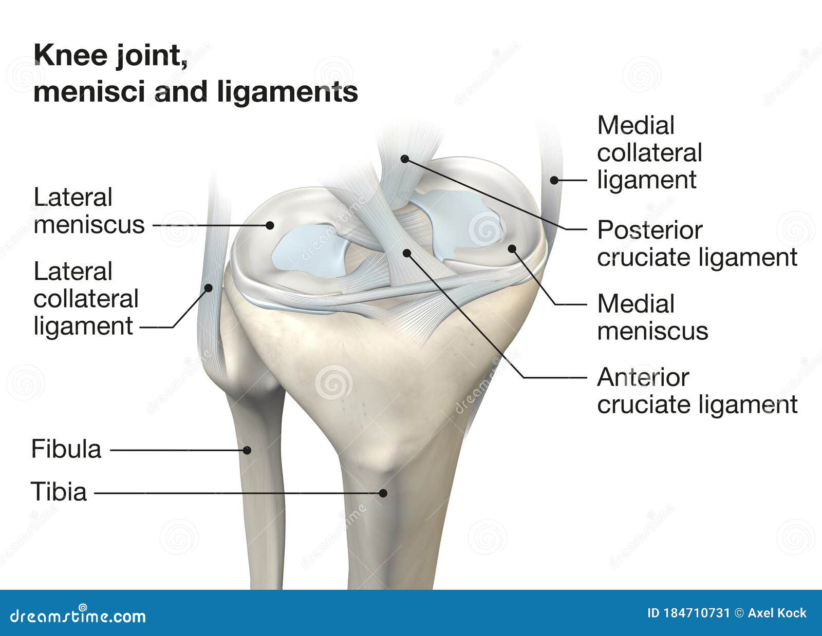
Knee Joint Anatomy Menisci And Ligaments Medically 3d Illustration The knee, also known as the tibiofemoral joint, is a synovial hinge joint formed between three bones: the femur, tibia, and patella. two rounded, convex processes (known as condyles) on the distal end of the femur meet two rounded, concave condyles at the proximal end of the tibia. the patella lies in front of the femur on the anterior surface. The knee joint is a synovial joint that connects three bones; the femur, tibia and patella. it is a complex hinge joint composed of two articulations; the tibiofemoral joint and patellofemoral joint. the tibiofemoral joint is an articulation between the tibia and the femur, while the patellofemoral joint is an articulation between the patella.

Knee Joint Anatomy Menisci And Ligaments Side View Medically 3d Vertices: 628.7k. more model information. noai: this model may not be used in datasets for, in the development of, or as inputs to generative ai programs. learn more. a model of the right knee showing some of the key bones, ligaments and tendons. published 5 years ago. science & technology 3d models. anatomy. Musculoskeletal system of the knee. in these topics. evaluation of the knee. knee pain. knee extensor mechanism injuries. brought to you by merck & co, inc., rahway, nj, usa (known as msd outside the us and canada) — dedicated to using leading edge science to save and improve lives around the world. learn more about the merck manuals and our. Part 1. this is a tutorial on the knee joint. the knee joint is the largest synovial joint in the body and it’s these articulations between the femur and the tibia and also between the patella and the femur. it’s a hinge joint and the main movements you get at this joint are flexion and extension. in this position here, which we’re. The knee joint is a hinge type synovial joint, which mainly allows for flexion and extension (and a small degree of medial and lateral rotation). it is formed by articulations between the patella, femur and tibia. in this article, we shall examine the anatomy of the knee joint – its articulating surfaces, ligaments and neurovascular supply.

Premium Photo 3d Illustration Of Human Body Knee Joint Anatomy Part 1. this is a tutorial on the knee joint. the knee joint is the largest synovial joint in the body and it’s these articulations between the femur and the tibia and also between the patella and the femur. it’s a hinge joint and the main movements you get at this joint are flexion and extension. in this position here, which we’re. The knee joint is a hinge type synovial joint, which mainly allows for flexion and extension (and a small degree of medial and lateral rotation). it is formed by articulations between the patella, femur and tibia. in this article, we shall examine the anatomy of the knee joint – its articulating surfaces, ligaments and neurovascular supply. Knee joint (art. genus) it connects three bones: the femur (femur), tibia (tibia) and patella (patella). this joint includes two separate joints. the first is the tibiofemoral joint, which is formed between the articular surfaces on the condyles of the femur (facies articularis condyli femoris) and the superior articular surface of the tibia. Knee anatomy involves more than just muscles and bones. ligaments, tendons, and cartilage work together to connect the thigh bone, shin bone, and knee cap and allow the leg to bend back and forth like a hinge. the largest joint in the body, the knee is also one of the most easily injured. problems with any part of the knee's anatomy can result.

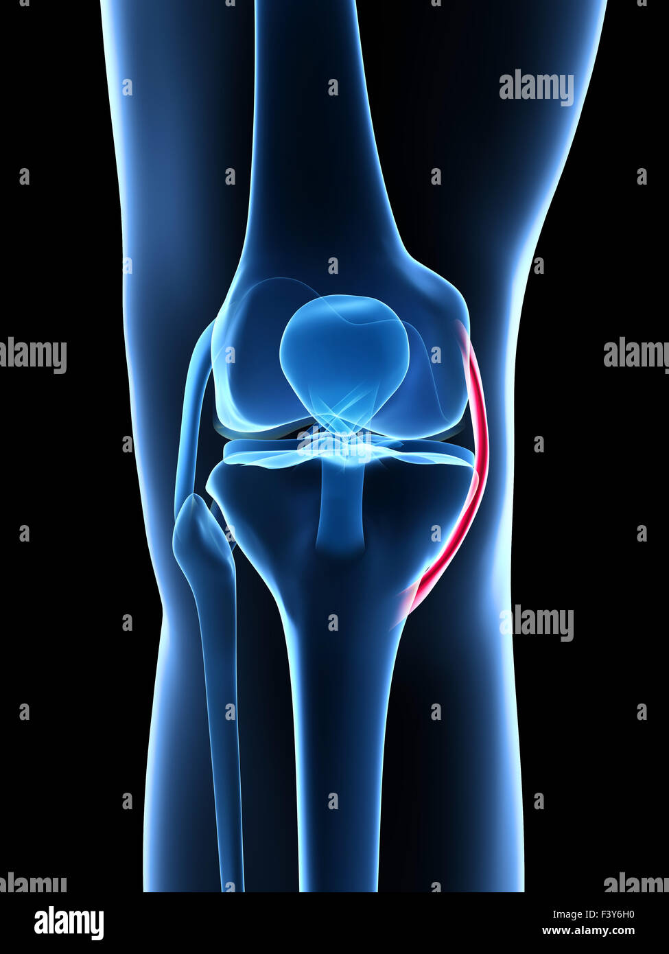
3d Rendered Illustration Knee Anatomy Stock Photo Alamy Knee joint (art. genus) it connects three bones: the femur (femur), tibia (tibia) and patella (patella). this joint includes two separate joints. the first is the tibiofemoral joint, which is formed between the articular surfaces on the condyles of the femur (facies articularis condyli femoris) and the superior articular surface of the tibia. Knee anatomy involves more than just muscles and bones. ligaments, tendons, and cartilage work together to connect the thigh bone, shin bone, and knee cap and allow the leg to bend back and forth like a hinge. the largest joint in the body, the knee is also one of the most easily injured. problems with any part of the knee's anatomy can result.

Comments are closed.