The Hyoid Bone Anatomy Function And Conditions
:max_bytes(150000):strip_icc()/GettyImages-670890563-5b6d0e6a46e0fb0050e8b985.jpg)
The Hyoid Bone Anatomy Function And Conditions Hyoid bone. your hyoid bone is in the front of your neck. it supports your tongue and plays a key role in speaking and swallowing. connected to nearby structures via ligaments, muscles and cartilage, your hyoid bone is the only “floating” bone in your body. contents overview function anatomy conditions and disorders care additional common. The anatomy of the hyoid bone. the hyoid bone is a small horseshoe shaped bone located in the front of your neck. it sits between the chin and the thyroid cartilage and is instrumental in the function of swallowing and tongue movements. the little talked about hyoid bone is a unique part of the human skeleton for a number of reasons.

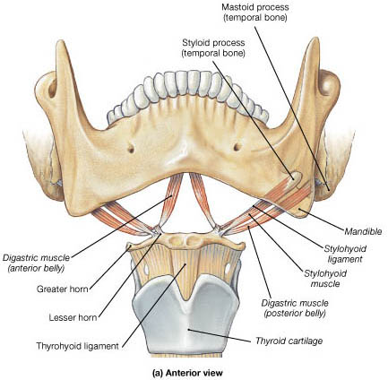
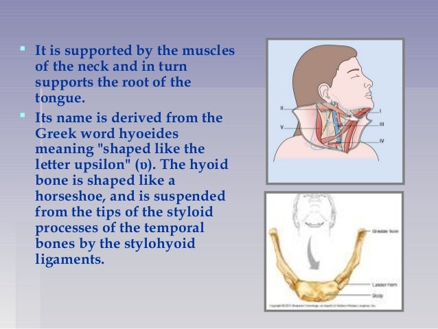
Hyoid Bone Description Anatomy Function Britannica Gross anatomy. the hyoid bone is a u shaped bone that is held in place by the strap muscles of the anterior triangle of the neck. the bone has a central body (forming the center of the “u”) with two smaller protruding structures on the superior surface (lesser horns) and two larger bony protrusions from the body (greater horns). Sternohyoid muscle: the muscle gets inserted in the inferior surface of the vocal bone. 2. omohyoid muscle: it inserts on the inferolateral surface of the vocal bone. 3. sternothyroid muscle: this muscle does not directly attach to the vocal bone. it inserts on the oblique line of the thyroid cartilage. 4. Typically, a broken hyoid results from forced strangulation (i.e. choking). the hyoid bone is located between the chin and the thyroid cartilage. it is also at the base of the mandible, or lower. The hyoid is composed of a body, two greater horns and two lesser horns: body – the central part of the bone. it has an anterior convex surface and a concave posterior surface. greater horn – projects from each end of the body in a posterior, superior and lateral direction. it acts as a site of attachment for numerous neck muscles.

Hyoid Bone Anatomy Function Anatomy Info Typically, a broken hyoid results from forced strangulation (i.e. choking). the hyoid bone is located between the chin and the thyroid cartilage. it is also at the base of the mandible, or lower. The hyoid is composed of a body, two greater horns and two lesser horns: body – the central part of the bone. it has an anterior convex surface and a concave posterior surface. greater horn – projects from each end of the body in a posterior, superior and lateral direction. it acts as a site of attachment for numerous neck muscles. Where is the hyoid bone located. the bone is located in the mid neck region, between the mandible and the thyroid cartilage, anterior to the pharynx and epiglottis at the level of the third cervical vertebra (c3). it remains suspended from the tips of the styloid processes of the temporal bones by the stylohyoid ligaments. The hyoid bone (hyoid) is a small u shaped (horseshoe shaped) solitary bone situated in the midline of the neck anteriorly at the base of the mandible and posteriorly at the fourth cervical vertebra. its anatomical position is just superior to the thyroid cartilage. it is closely linked with an extended tendon muscular complex but not specifically interconnected to any adjacent bones and.

Anatomy And Functions Of Hyoid Where is the hyoid bone located. the bone is located in the mid neck region, between the mandible and the thyroid cartilage, anterior to the pharynx and epiglottis at the level of the third cervical vertebra (c3). it remains suspended from the tips of the styloid processes of the temporal bones by the stylohyoid ligaments. The hyoid bone (hyoid) is a small u shaped (horseshoe shaped) solitary bone situated in the midline of the neck anteriorly at the base of the mandible and posteriorly at the fourth cervical vertebra. its anatomical position is just superior to the thyroid cartilage. it is closely linked with an extended tendon muscular complex but not specifically interconnected to any adjacent bones and.

Comments are closed.