The Anatomy Of Elbow Joint Powerpoint Presentation

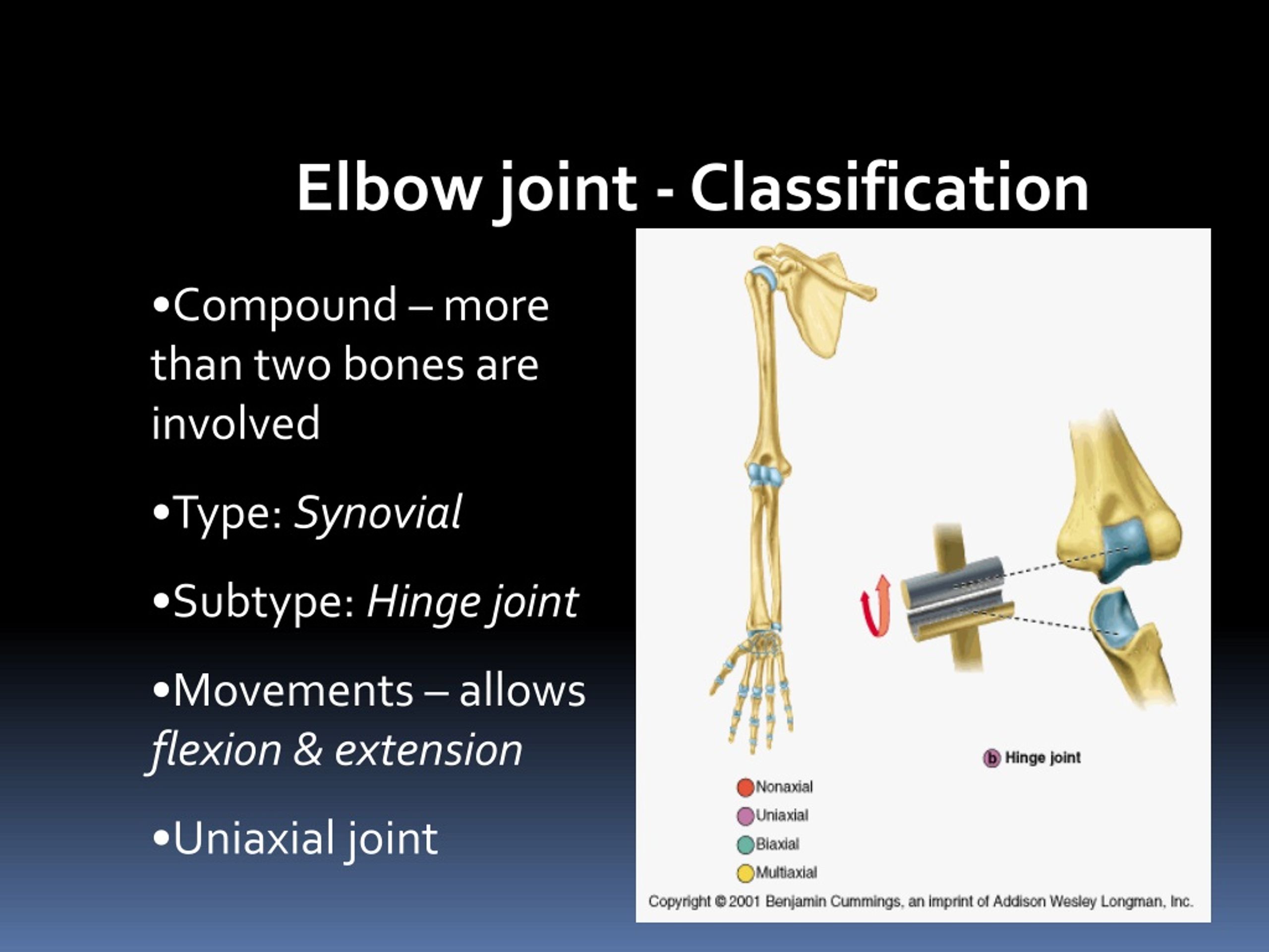
Ppt The Elbow Powerpoint Presentation Free Download Id 2188501 Anatomy of elbow joint. the elbow joint is formed by three joints: the humeroulnar joint, humeroradial joint, and proximal radio ulnar joint. it is a synovial hinge joint that allows movement in one plane. the trochlea and capitellum are the proximal articular surfaces that form joints with the ulna and radius. The elbow joint is formed by three joints: the humeroulnar joint, humeroradial joint, and proximal radio ulnar joint. it is a synovial hinge joint that allows movement in one plane. the trochlea and capitellum are the proximal articular surfaces that form joints with the ulna and radius. distally, the head of the radius and olecranon process of.

Ppt Elbow Joint Powerpoint Presentation Free Download Id 8796306 3. elbow joint. the document summarizes the anatomy of the elbow joint, proximal radioulnar joint, and distal radioulnar joint. it describes the articulating bones, ligaments, capsules, synovial membranes, and movements of flexion, extension, pronation, and supination at each joint. key points are that the elbow is a hinge joint that flexes and. Elbow anatomy and biomechanics . mimi renaudin, dpt university of mississippi medical center. objectives. describe the anatomy and joint articulations at the elbow discuss the static and dynamic constraints acting at the elbow identify the neurovascular contributions within the elbow joint. download presentation. Download presentation. presentation on theme: "elbow joint."—. presentation transcript: 1 elbow joint. 2 elbow anatomy elbow joint is made of 3 bones 2 joints one capsule. hinge joint flexion (145) and extension. 3 elbow anatomy elbow joint where the radius and ulna articulate with the humerus. flexion and extension hinge joint. Dr rania gabr. elbow join t. type: uniaxial, synovial hinge joint articulation : trochlea and capitulum of the humerus above trochlear notch of ulna and the head of radius below the articular surfaces are covered with articular (hyaline) cartilage. capitulum. download presentation. articular surfaces. dislocations fractures. collagenous tissues.

Ppt Elbow Joint Powerpoint Presentation Free Download Id 8796306 Download presentation. presentation on theme: "elbow joint."—. presentation transcript: 1 elbow joint. 2 elbow anatomy elbow joint is made of 3 bones 2 joints one capsule. hinge joint flexion (145) and extension. 3 elbow anatomy elbow joint where the radius and ulna articulate with the humerus. flexion and extension hinge joint. Dr rania gabr. elbow join t. type: uniaxial, synovial hinge joint articulation : trochlea and capitulum of the humerus above trochlear notch of ulna and the head of radius below the articular surfaces are covered with articular (hyaline) cartilage. capitulum. download presentation. articular surfaces. dislocations fractures. collagenous tissues. 1 5. synonyms: none. the elbow joint is a synovial joint found in the upper limb between the arm and the forearm. it is the point of articulation of three bones: the humerus of the arm and the radius and the ulna of the forearm. the elbow joint is classified structurally as a synovial joint. it is also classified structurally as a compound. Slide 25 . stress tests valgus (ucl) varus (rcl) stress test – positive sign is laxity tennis elbow test (lateral epicondylitis) positive sign is increased pain tinel’s sign (ulnar nerve) – numbness, tinkling into ulnar nerve region. free download elbow anatomy powerpoint presentation.

Comments are closed.