Spinal Cord Anatomy V3 0
:watermark(/images/watermark_5000_10percent.png,0,0,0):watermark(/images/logo_url.png,-10,-10,0):format(jpeg)/images/overview_image/1099/FtvR9dbol9TTwZl8o97pDQ_anatomy-vertebral-column_english.jpg)
Spinal Cord Anatomy Structure Tracts And Function Kenhub Brief overview of spinal cord anatomy. The spinal cord is part of the central nervous system (cns). it is situated inside the vertebral canal of the vertebral column. during development, there’s a disproportion between spinal cord growth and vertebral column growth. the spinal cord finishes growing at the age of 4, while the vertebral column finishes growing at age 14 18.

Spinal Cord Anatomy Anatomy Reading Source Csf is an ultrafiltrate of blood plasma through the permeable capillaries of the choroid plexus. volume. total csf volume between brain, spinal cord, and thecal sac is ~150 ml. csf formation occurs at rate of ~500 ml per day. thus, the total amount of csf is turned over 3 4 times per day. nerve root anatomy. cervical spine. The vertebra provides several crucial functions to the body. first, it acts as a structural component of the spine, bearing body weight, anchoring muscles and the spinal cord, and forming joints with other vertebrae and ribs that allow the torso and neck to move. second, the vertebra protects the delicate tissues of the spinal cord by. The spinal cord is a tubular bundle of nervous tissue and supporting cells that extends from the brainstem to the lumbar vertebrae. together, the spinal cord and the brain form the central nervous system. in this article, we shall examine the macroscopic anatomy of the spinal cord – its structure, membranous coverings and blood supply. An essential feature of the central nervous system (cns), the spinal cord lies within the spinal column and extends from the brainstem to the lower back through the vertebral foramen of the vertebrae. in adults, the spinal cord terminates in the lumbar region at l1 l2, the conus medullaris.[1] below this, the vertebral canal contains the "cauda equina" or "horse's tail," a bundle of nerve roots.

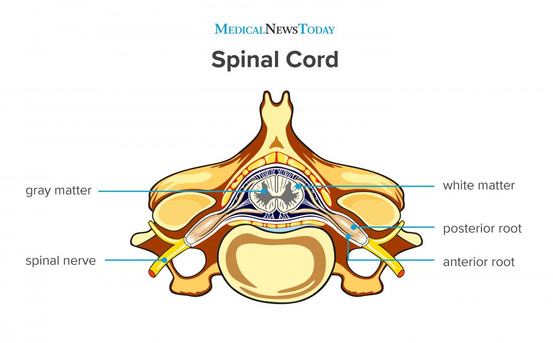
Spinal Cord Labeled The spinal cord is a tubular bundle of nervous tissue and supporting cells that extends from the brainstem to the lumbar vertebrae. together, the spinal cord and the brain form the central nervous system. in this article, we shall examine the macroscopic anatomy of the spinal cord – its structure, membranous coverings and blood supply. An essential feature of the central nervous system (cns), the spinal cord lies within the spinal column and extends from the brainstem to the lower back through the vertebral foramen of the vertebrae. in adults, the spinal cord terminates in the lumbar region at l1 l2, the conus medullaris.[1] below this, the vertebral canal contains the "cauda equina" or "horse's tail," a bundle of nerve roots. Anatomy. the spinal cord is a mass of nervous tissue that extends inferiorly from the brain stem through the vertebral canal of the cervical and thoracic regions, ending around the t12 or l1 vertebra. it is a long tube about 18 inches (45 cm) in length and around half an inch (1 cm) in diameter at its widest point. The spinal cord is highly organized into different segments and contains both gray and white matter, each with distinct roles. below is a detailed description of its anatomy: gross structure. the spinal cord is approximately 42 45 cm long in adults and has a diameter of about 1 cm.

Spinal Cord Anatomy Physiopedia Anatomy. the spinal cord is a mass of nervous tissue that extends inferiorly from the brain stem through the vertebral canal of the cervical and thoracic regions, ending around the t12 or l1 vertebra. it is a long tube about 18 inches (45 cm) in length and around half an inch (1 cm) in diameter at its widest point. The spinal cord is highly organized into different segments and contains both gray and white matter, each with distinct roles. below is a detailed description of its anatomy: gross structure. the spinal cord is approximately 42 45 cm long in adults and has a diameter of about 1 cm.

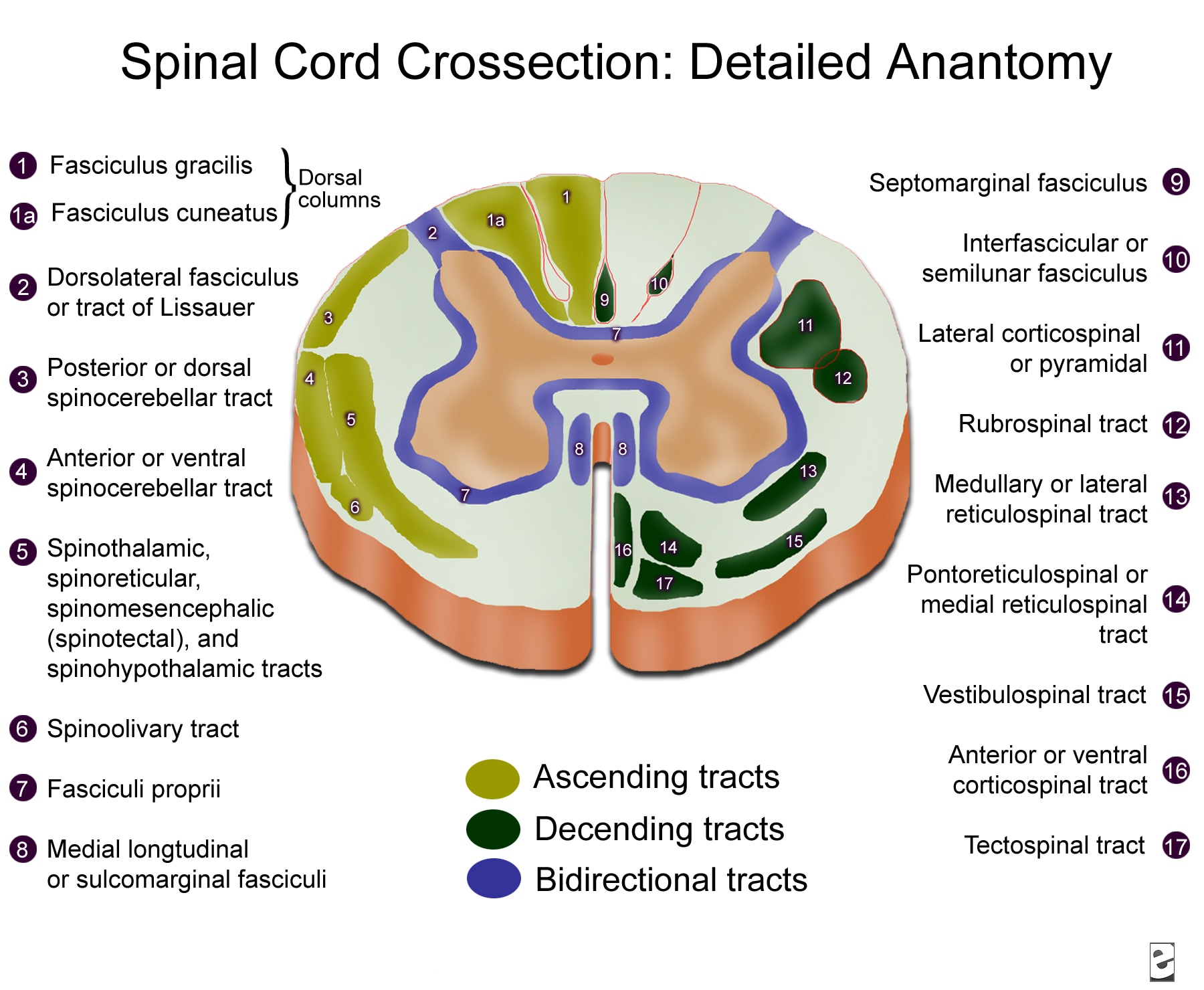
Spinal Cord Cross Sectional Anatomy R Diology De Arun

Comments are closed.