Second Trimester Transvaginal Scan And Three Dimensional Ultrasound

Second Trimester Transvaginal Scan And Three Dimensional Ultrasound Continuing education activity. the mid trimester obstetric ultrasound (also known as the second trimester anatomy scan or anomaly scan) is a routine examination in many countries. it plays a pivotal role in ensuring the well being of both the pregnant patient and the developing fetus. a cornerstone of modern prenatal care, this non invasive. Historically the second trimester ultrasound was often the only routine scan offered in a pregnancy and so was expected to provide information about gestational age (correcting menstrual dates if necessary), fetal number and type of multiple pregnancy, placental position and pathology, as well as detecting fetal abnormalities. 1 many patients.

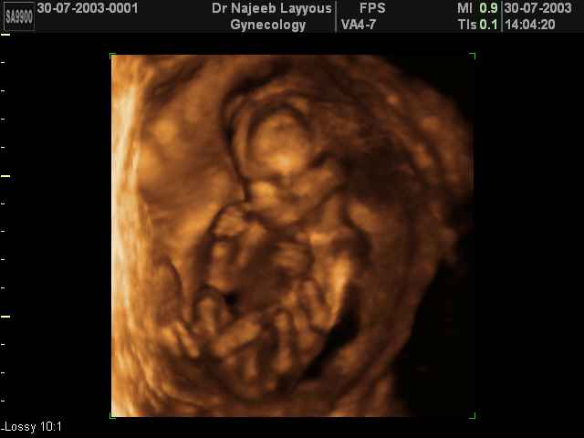
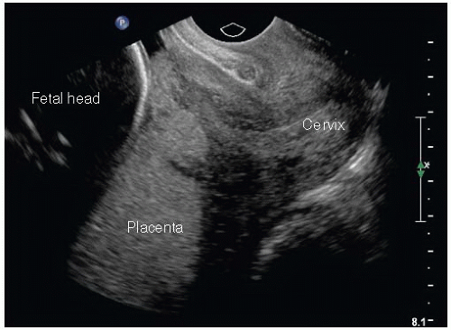
Second Trimester Transvaginal Scan And Three Dimensional Ultrasound Introduction. the second trimester ultrasound is commonly performed between 18 and 22 weeks gestation. historically the second trimester ultrasound was often the only routine scan offered in a pregnancy and so was expected to provide information about gestational age (correcting menstrual dates if necessary), fetal number and type of multiple pregnancy, placental position and pathology, as. Second trimester transvaginal scan and three dimensional ultrasound with “glass body” rendering (a), doppler ultrasound (b) and two dimensional ultrasound showing the anatomical relationship between the cervical canal (yellow lines) (c) and the presence of vasa praevia (red arrow) (d). figure 17. Evaluation for abnormalities such as fibroids, adnexal masses, mullerian duct anomalies is evaluated in the first trimester and mid second trimester scans. the presence of ultrasound abnormalities or clinical indications can lead to additional specialized ultrasound examinations such as serial transvaginal cervical lengths or limited follow up. Descriptions of mbhb development on transvaginal sonography (tvs) have been published mostly for the second half of pregnancy and include reference ranges for the fetal vermis, brainstem (bs), pons and the cerebral aqueduct (aq) 1 3. leibovitz et al. 4 described normative reference ranges for various mbhb components using three dimensional (3d.

3d Second Trimester Ultrasound Scan Photos Second Part Of Pregnancy Evaluation for abnormalities such as fibroids, adnexal masses, mullerian duct anomalies is evaluated in the first trimester and mid second trimester scans. the presence of ultrasound abnormalities or clinical indications can lead to additional specialized ultrasound examinations such as serial transvaginal cervical lengths or limited follow up. Descriptions of mbhb development on transvaginal sonography (tvs) have been published mostly for the second half of pregnancy and include reference ranges for the fetal vermis, brainstem (bs), pons and the cerebral aqueduct (aq) 1 3. leibovitz et al. 4 described normative reference ranges for various mbhb components using three dimensional (3d. Ultrasound is generally considered safe during pregnancy [5]. it should, however, only be performed for a valid medical indication [2]. the lowest possible energy and time exposure should be used to obtain necessary information [1]. protocol for the second trimester anomalies scan the second trimester scan includes three components. Three dimensional volumes were acquired during the second trimester of pregnancy, between 11 and 24 weeks of gestation, using a standardized protocol. the participants were examined in the presence of a chaperone and with consent, in supine gynecological position, post micturition.

Second And Third Trimester Pregnancy Radiology Key Ultrasound is generally considered safe during pregnancy [5]. it should, however, only be performed for a valid medical indication [2]. the lowest possible energy and time exposure should be used to obtain necessary information [1]. protocol for the second trimester anomalies scan the second trimester scan includes three components. Three dimensional volumes were acquired during the second trimester of pregnancy, between 11 and 24 weeks of gestation, using a standardized protocol. the participants were examined in the presence of a chaperone and with consent, in supine gynecological position, post micturition.

Three Dimensional Transvaginal Ultrasound Depicting Multiplanar Display

Comparison Of Two Dimensional Transvaginal Ultrasound Three

Comments are closed.