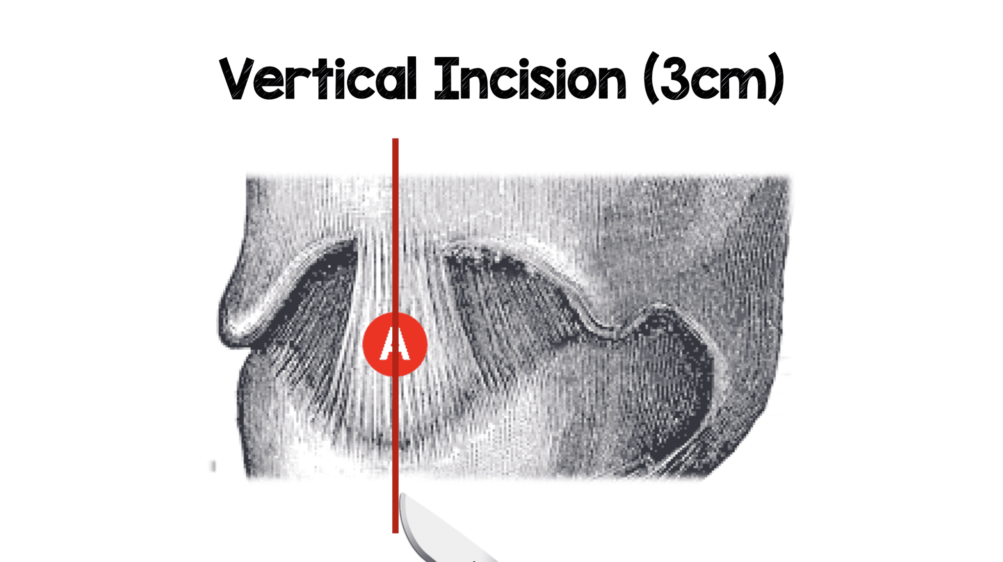
Schematic Illustration Of The Surgical Technique A Vertical Incision

Schematic Illustration Of The Surgical Technique A Vertical Incision Schematic illustration of the surgical technique (a) vertical incision of the orbicularis oris muscle flaps was performed. dissection of the muscle flap was performed at the gentian violet markings. Introduction. there are now thousands of randomized trials on the different steps for the technique for cesarean birth. the following discussion will review each step in the procedure and provide evidence based recommendations for surgical technique, when these data are available. in many cases, when comparisons showed statistical significance.

Suturing Of Surgical Incision Illustration Stock Image C046 3002 Fig. 5. extraction of the fetal head. the surgeon's dominant hand is placed into the uterine incision so that the back of the hand is against the inside of the lower uterine segment and the fingers cup the fetal head. firm, gentle traction is used to elevate the fetal head toward the incision. Through the vertical incision, uterine closure is technically difficult. to decrease the risks of hemorrhage and adhesion, a speedy and skillful technique is mandatory. the most serious risk of vertical incision in the contractile corpus is uterine rupture in the subsequent pregnancy. therefore, cases of prior classical cesarean section are. Interestingly, the vertical opening of the abdomen was still the main technique used in the 1970s, although it was known from the beginning of the twentieth century to be associated with higher rates of long term postoperative complications such as wound dehiscence and abdominal incision hernia and cosmetic issues compared to the transverse. A transverse incision is associated with less postoperative pain, greater wound strength, and better cosmetic results than the vertical midline incision. 13, 14 vertical incisions generally allow faster abdominal entry, cause less bleeding and nerve injury, and can be easily extended in a cephalad direction if more space is required for access.

Schematic Diagram Of Operation A Surgical Incision The Incision Interestingly, the vertical opening of the abdomen was still the main technique used in the 1970s, although it was known from the beginning of the twentieth century to be associated with higher rates of long term postoperative complications such as wound dehiscence and abdominal incision hernia and cosmetic issues compared to the transverse. A transverse incision is associated with less postoperative pain, greater wound strength, and better cosmetic results than the vertical midline incision. 13, 14 vertical incisions generally allow faster abdominal entry, cause less bleeding and nerve injury, and can be easily extended in a cephalad direction if more space is required for access. Schematic illustration of the eight different types of neck incisions. open in a new tab (a) midline vertical incision, (b) t shaped incision, (c) horizontal double y incision, (d) trap door incision, (e) double trap doors, (f) apron incision (separate tracheostoma), (g) apron incision (tracheostoma incorporated), and (h) extended apron incision. Figure 3. the skin is fully incised. in the joel cohen technique, the skin incision is placed 3 cm above the original pfannenstiel incision, the subcutaneous tissue is incised only in the three most medial centimetres, and the lateral tissue is separated manually, before the fascia is divided bluntly with both index fingers inserted in the deep fascial space created by the knife.

Vertical Incision Rebel Em Emergency Medicine Blog Schematic illustration of the eight different types of neck incisions. open in a new tab (a) midline vertical incision, (b) t shaped incision, (c) horizontal double y incision, (d) trap door incision, (e) double trap doors, (f) apron incision (separate tracheostoma), (g) apron incision (tracheostoma incorporated), and (h) extended apron incision. Figure 3. the skin is fully incised. in the joel cohen technique, the skin incision is placed 3 cm above the original pfannenstiel incision, the subcutaneous tissue is incised only in the three most medial centimetres, and the lateral tissue is separated manually, before the fascia is divided bluntly with both index fingers inserted in the deep fascial space created by the knife.

Comments are closed.