Pocus Made Easy Lung Litfl Ultrasound Library

Pocus Made Easy Lung Litfl Ultrasound Library Labelled right lat inf. assess lung diaphragm interface (more sensitive for dependent lung pathology) 5 to 8. same as above on left chest *left. image sets. minimum 10 video loops. 8x lung field (4 each side) 2x pleural assessment (1 each side) 10 14 video loops 2 images. Welcome to the pocus made easy pages! below are 11 short teaching videos (2 8 minutes) and educational pages summarising how to perform the following emergency department pocus exams. these resources are designed with simplicity and ed specific ultrasound goals in mind. you can use these resources to augment your knowledge or revise pocus exam.

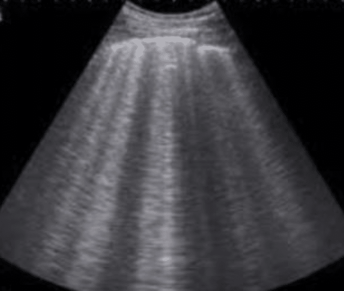
Pocus Made Easy Lung Litfl Ultrasound Library Pocus made easy: efast. fahad yousif. aug 21, 2023. home ultrasound library. efast (focused assessment with sonography in trauma) is used to look for pathology in trauma such as haemothorax, pneumothorax, haemoperitoneum and haemopericardium and looking for free fluid in ectopic pregnancy. pre reading. Place your probe at the right (r2) or left (l2) midaxillary line around the 6 7th intercostal space. this should be just lateral to the nipple line in males. anchor your probe between two ribs, and just like in point 1, look for the batwing sign, lung sliding, and a lines. r2 ultrasound probe position. Place your probe at the mid clavicular line at the 2nd intercostal space of the right and left lungs respectively. this point is the most sensitive spot for looking for pneumothorax in the supine patient. anchor your probe in the space between two ribs and set the ultrasound machine depth to 3 5cm. Lung ultrasound. this infographic explores artifacts in point of care ultrasound (pocus) to aid diagnosis when performing a ultrasound on the lungs. note the differences between the a lines and b lines scans. download the pdf a lines multiple repeating horizontal lines that are parallel and equidistant from the pleura. this can be a […].

Pocus Made Easy Lung Litfl Ultrasound Library Place your probe at the mid clavicular line at the 2nd intercostal space of the right and left lungs respectively. this point is the most sensitive spot for looking for pneumothorax in the supine patient. anchor your probe in the space between two ribs and set the ultrasound machine depth to 3 5cm. Lung ultrasound. this infographic explores artifacts in point of care ultrasound (pocus) to aid diagnosis when performing a ultrasound on the lungs. note the differences between the a lines and b lines scans. download the pdf a lines multiple repeating horizontal lines that are parallel and equidistant from the pleura. this can be a […]. Thoracic ultrasound has rapidly gained popularity over the past 10 years, mainly due to its wide availability in emergency and trauma settings, as well as trends toward the point of care ultrasound (pocus) usage in training programs. additionally, ultrasound circumvents many of the issues that arrive with traditional radiography, such as delay of care and radiation exposure. in an unstable. Surprisingly, lung ultrasound had a much higher sensitivity of 92% and a specificity of 99.4%. ct scan of the chest had the highest sensitivity and specificity out of all the modalities. the study concluded that lung ultrasound performed by trained users in the emergency department to detect occult pneumothorax was almost as accurate as ct scan.

Pocus Made Easy Efast Litfl Ultrasound Library Thoracic ultrasound has rapidly gained popularity over the past 10 years, mainly due to its wide availability in emergency and trauma settings, as well as trends toward the point of care ultrasound (pocus) usage in training programs. additionally, ultrasound circumvents many of the issues that arrive with traditional radiography, such as delay of care and radiation exposure. in an unstable. Surprisingly, lung ultrasound had a much higher sensitivity of 92% and a specificity of 99.4%. ct scan of the chest had the highest sensitivity and specificity out of all the modalities. the study concluded that lung ultrasound performed by trained users in the emergency department to detect occult pneumothorax was almost as accurate as ct scan.

Comments are closed.