Pelvic Anatomy Female Ligaments

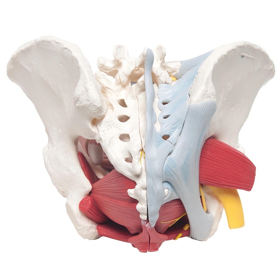
Anatomical Models Of Female Pelvis With Ligaments Vessels Nerves The ligaments of the female reproductive tract are a series of structures that support the internal female genitalia in the pelvis. the ligaments of the female reproductive tract can be divided into three categories: broad ligament – a sheet of peritoneum, associated with both the uterus and ovaries. uterine ligaments – ligaments primarily. The pelvis contains a large number of organs, bones, muscles, and ligaments, so many conditions can affect the entire pelvis or parts within it. some conditions that can affect the female pelvis.

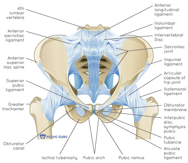
Representative Image Of The Pelvic Floor Ligaments And Their Relative The anatomy of the female genital tract and lower urinary and gastrointestinal tracts relevant to the surgeon performing laparotomy or laparoscopy, with an emphasis on clinical relevance and avoiding potential complications, is reviewed here. surgical pelvic anatomy from a vaginal approach and the surgical anatomy of the anterior abdominal wall. The pelvis's bony integrity is supported by various ligaments that lend crucial flexible strength to the pelvic cavity and support some of the internal pelvic structures.[1] the principal ligaments are sacrotuberous, sacrospinous, and iliolumbar, and in the female pelvis, there are further ligaments to support the ovaries and uterus (see image. pelvic ligaments).[2] trauma or stretching of. Imagine the vagina and uterus forming a slightly tilted capital p in a sagittal section of the female pelvis. this organ is divided into three parts, the body, the isthmus and the cervix, which are supported by several ligaments and pelvic muscles. ligaments of the uterus. in this discussion, a "ligament" refers. Structure and function. the broad ligament functions as a protective layer for the female pelvic organs. it carries blood vessels, nerves, and lymphatics to the structures within the mesentery. the broad ligament consists of a double layer of peritoneum, and the different parts are named based on the structures contained between the double layer.

Surgical Exposure And Anatomy Of The Female Pelvis Surgical Clinics Imagine the vagina and uterus forming a slightly tilted capital p in a sagittal section of the female pelvis. this organ is divided into three parts, the body, the isthmus and the cervix, which are supported by several ligaments and pelvic muscles. ligaments of the uterus. in this discussion, a "ligament" refers. Structure and function. the broad ligament functions as a protective layer for the female pelvic organs. it carries blood vessels, nerves, and lymphatics to the structures within the mesentery. the broad ligament consists of a double layer of peritoneum, and the different parts are named based on the structures contained between the double layer. The female reproductive system is made up of external and internal organs. the external organs lie in an area called the vulva, and they include the labia, the clitoris, and the vaginal opening. the internal reproductive organs can be found within the pelvic cavity, and they include the ovaries, which produce the female sex cells, called. The bony pelvis is a complex basin shaped structure that comprises the skeletal framework of the pelvic region and houses the pelvic organs. it consists of the hip bone and the sacrum, which are connected via the sacroiliac joint. the hip bone is composed of three fused bones: the ilium, ischium and the pubic bone.

What Is The Pelvic Floor Your Pace Yoga The female reproductive system is made up of external and internal organs. the external organs lie in an area called the vulva, and they include the labia, the clitoris, and the vaginal opening. the internal reproductive organs can be found within the pelvic cavity, and they include the ovaries, which produce the female sex cells, called. The bony pelvis is a complex basin shaped structure that comprises the skeletal framework of the pelvic region and houses the pelvic organs. it consists of the hip bone and the sacrum, which are connected via the sacroiliac joint. the hip bone is composed of three fused bones: the ilium, ischium and the pubic bone.

Comments are closed.