Pectoral Region Muscles Anatomy Earth S Lab

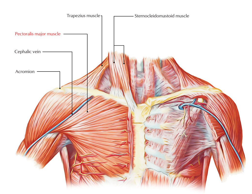
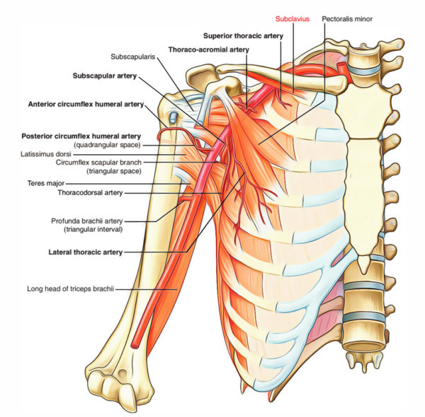
Pectoral Region Muscles Anatomy Earth S Lab The pectoralis minor is regarded as the ‘key muscle’ of axilla because it crosses in front of the axillary artery and thus used to split this artery into three parts. the origin of pectoralis minor is varied. it may be prefixed (i.e., arises from 2nd to 5th ribs) or postfixed (i.e., arises from 4th to 6th ribs). Contents. the pectoralis major is a large, broad, fan shaped muscle located on the front side of the thorax. it is the biggest muscle of the pectoral region. it travels across the front of the underarm, where it stays thick, and afterwards connects to its slim, square tendon of attachment, which allows it to enter underneath the deltoid towards.

Pectoral Region Muscles Anatomy Earth S Lab Clavipectoral fascia. the clavipectoral fascia is a strong fascial sheet deep to the clavicular head of the pectoralis major muscle, filling the space between the clavicle and the pectoralis minor muscle. a as observed in sagittal section of anterior axillary wall; b as viewed from front. The pectoral region is located on the anterior chest wall. it contains four muscles that exert a force on the upper limb: the pectoralis major, pectoralis minor, serratus anterior and subclavius. in this article, we shall look at the anatomy of the muscles of the pectoral region – their attachments, actions and innervation. Comprehensive anatomy of the pectoral region including origin, course, and distribution of the major vessels and their branches that supply drain it. muscle innervations from the brachial plexus, together with their sensory distribution to predict or localize the consequences of injury to these nerves along with an explanation on how to test their functional integrity clinically. Terms in this set (7) what are the 3 muscles of the pectoral region? pectoralis major, minor, serratus anterior. what are the pectoralis major actions? adduction, medially rotate, & flex shoulder. what are pectoralis major innervations? medial pectoral n. and lateral pectoral n.(medial pectoral n. of medial cord, lateral pectoral n. off of.

Pectoral Region Muscles Anatomy Earth S Lab Comprehensive anatomy of the pectoral region including origin, course, and distribution of the major vessels and their branches that supply drain it. muscle innervations from the brachial plexus, together with their sensory distribution to predict or localize the consequences of injury to these nerves along with an explanation on how to test their functional integrity clinically. Terms in this set (7) what are the 3 muscles of the pectoral region? pectoralis major, minor, serratus anterior. what are the pectoralis major actions? adduction, medially rotate, & flex shoulder. what are pectoralis major innervations? medial pectoral n. and lateral pectoral n.(medial pectoral n. of medial cord, lateral pectoral n. off of. 1 4. synonyms: none. the pectoralis major muscle is a broad superficial muscle found superficially in the anterior chest wall. in males, it is covered by the deep layer of fascia, subcutaneous tissue and the adjacent skin. in females, it is covered by the breast. the deep surface of the muscle covers the pectoralis minor and serratus anterior. What is the pectoralis major. the pectoralis major is a large, triangular, or fan shaped superficial muscle lying at the anteriormost position in the chest cavity. it forms the front wall of the axilla, making up most of the chest and breast. the paired muscle is often called the ‘pecs’ in combination with the smaller muscle pectoralis minor.

Anatomy Of Pectoral Region 1 4. synonyms: none. the pectoralis major muscle is a broad superficial muscle found superficially in the anterior chest wall. in males, it is covered by the deep layer of fascia, subcutaneous tissue and the adjacent skin. in females, it is covered by the breast. the deep surface of the muscle covers the pectoralis minor and serratus anterior. What is the pectoralis major. the pectoralis major is a large, triangular, or fan shaped superficial muscle lying at the anteriormost position in the chest cavity. it forms the front wall of the axilla, making up most of the chest and breast. the paired muscle is often called the ‘pecs’ in combination with the smaller muscle pectoralis minor.

Solution Anatomy Of The Upper Limb Lab The Pectoral Region Studypool

Pectoral Region Lab Yed聴tepe Anatomy Lab

Comments are closed.