Nerve Tissue Stain Pathologists Medicallaboratorytechnology Nimhans Neuropathy Mbbs Medical

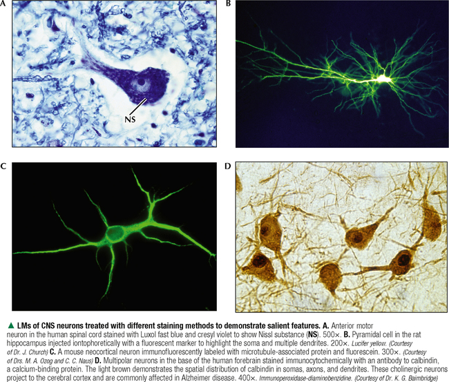
Nervous Tissue Basicmedical Key Confocal images of βiii tubulin staining dermal nerve bundles. (a) a neurovascular bundle with a nerve bundle (arrow) stained by βiii tubulin (green) and small branching fibers innervating a blood vessel (red). scale bar = 20 μm. (b) a dermal nerve bundle (arrows) stained by βiii tubulin (green). schwann cell nuclei are blue. Abstract. nerve biopsy is a valuable tool in the diagnostic work up of peripheral neuropathies. currently, major indications include interstitial pathologies such as suspected vasculitis and amyloidosis, atypical cases of inflammatory neuropathy and the differential diagnosis of hereditary neuropathies that cannot be specified otherwise.

Chapter 7 Nervous Tissue Histology An Identification Manual Nerve biopsy may then be very helpful in disclosing inflammation or other important interstitial pathology such as amyloidosis or immunoglobulin deposits associated with dysproteinemic neuropathy. it may also give clues to the underlying gene defect in unsolved cases of familial neuropathy, and may reveal combined pathology, for example. Neuromuscular pathology. section menu. the johns hopkins department of neurology offers comprehensive services for the evaluation of nerve and muscle disease, including: clinical evaluation. nerve and muscle biopsy. tissue preparation. pathological interpretation. nerve and muscle biopsies are performed by specially trained physicians, usually. The peripheral nerve society has proposed consensus guidelines for the diagnosis of definitive vasculitic neuropathy, probable vasculitic neuropathy, and possible vasculitic neuropathy that are briefly summarized below . these rely in part on a clear distinction between perivascular inflammation and inflammation within vessel walls. This pathology may be the result of metabolic, toxic, immune mediated, and or genetic factors. small fiber symptoms can be variable and inconsistent and therefore require an objective biomarker confirmation. small fiber dysfunction is not typically captured by diagnostic tests for large fiber neuropathy (nerve conduction and electromyographic.

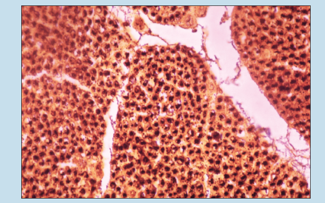
Light Microscopy Images Of Nerves H E Staining A Untreated Peripheral The peripheral nerve society has proposed consensus guidelines for the diagnosis of definitive vasculitic neuropathy, probable vasculitic neuropathy, and possible vasculitic neuropathy that are briefly summarized below . these rely in part on a clear distinction between perivascular inflammation and inflammation within vessel walls. This pathology may be the result of metabolic, toxic, immune mediated, and or genetic factors. small fiber symptoms can be variable and inconsistent and therefore require an objective biomarker confirmation. small fiber dysfunction is not typically captured by diagnostic tests for large fiber neuropathy (nerve conduction and electromyographic. Histological method as quality control in pnte. longitudinal section of a peripheral nerve stained with masson's trichrome method (a). transversal section of a neuragen ® collagen conduit stained with the mcoll histochemical method with low (b) and high magnification (c), where it is possible to observe the collagen fibers in red, the myelin in blue and the nucleus darkly stained. The approach to the neuropathological assessment of nerve biopsies is the main focus of this review. nerve biopsies are invasive diagnostic procedures resulting in a permanent neurological deficit, and are therefore carried out only following an in depth clinical assessment including laboratory, imaging, electrophysiological, and where appro.

Comments are closed.