Microkeratome Lasik Centre Ophtalmologique Agora

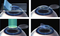
Microkeratome Lasik Centre Ophtalmologique Agora Microkeratome lasik centre ophtalmologique agora. accueil; centre ophtalmologique agora les cabinets ophtalmologiques 2 centres neufs, modernes, facilement. Cette technique se décompose en quatre étapes : 1. la découpe d’un volet cornéen superficiel ( épaisseur entre 90 et 120µm) 2. on soulève le volet cornéen pour exposer le stroma cornéen. 3. traitement par laser excimer du défaut de vision : myopie, hypermétropie, astigmatisme et presbytie. 4.

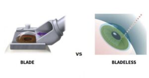
Microkeratome Lasik With Mel 90 Youtube Thus, immunohistological staining for specific inflammatory cells shows conclusively more monocytes in the cornea after formation of flaps with a femtosecond laser than with a microkeratome. when side cut energies are decreased, there is less epithelial injury and lower release of these proinflammatory cytokines and inflammation. Results. corneal backscatter was 6% higher after bladeless lasik than after lasik with the mechanical microkeratome at 1 month (p = 0.007), but not at 3 or 6 months. high contrast visual acuity, contrast sensitivity, and forward light scatter did not differ between treatments at any examination. flap thicknesses at 1 month were 143±16 μm. This article discusses the clinical advantages and disadvantages of the femtosecond laser and mechanical microkeratome for lasik flap creation. mechanical microkeratomes mechanical microkeratome devices (figure 1) use high precision oscillating blade systems that dock to a suction ring to create a lamellar corneal flap while the cornea is held. Laser assisted in situ keratomileusis (lasik) with a mechanical microkeratome compared to lasik with a femtosecond laser for lasik in adults with myopia or myopic astigmatism. kahuam lópez n, navas a, castillo salgado c, graue hernandez eo, jimenez corona a, ibarra a. cochrane database syst rev. 2020 apr 7; 4(4):cd012946.

Microkeratome Lasik Meaning How It Works Benefits And Risks This article discusses the clinical advantages and disadvantages of the femtosecond laser and mechanical microkeratome for lasik flap creation. mechanical microkeratomes mechanical microkeratome devices (figure 1) use high precision oscillating blade systems that dock to a suction ring to create a lamellar corneal flap while the cornea is held. Laser assisted in situ keratomileusis (lasik) with a mechanical microkeratome compared to lasik with a femtosecond laser for lasik in adults with myopia or myopic astigmatism. kahuam lópez n, navas a, castillo salgado c, graue hernandez eo, jimenez corona a, ibarra a. cochrane database syst rev. 2020 apr 7; 4(4):cd012946. Microkeratome accuracy in lasik. dennis s.c lam ∙ srinivas k rao ∙ king yu liu. download pdf. dear editor. the recent article by durairaj et al, concurs with earlier reports 1–3 that currently available microkeratomes create corneal flaps that are thinner than intended. these studies also indicate that variability in flap thickness is. Figure 2 confocal microscopy images (confoscan, nidek technologies) of both corneas of 1 subject before and after lasik. the right eye was randomized to bladeless lasik, and the left eye to lasik with the mechanical microkeratome. preoperative images are from a depth equivalent to the postoperative interface and show normal keratocyte nuclei.

Lasik Microkeratome Youtube Microkeratome accuracy in lasik. dennis s.c lam ∙ srinivas k rao ∙ king yu liu. download pdf. dear editor. the recent article by durairaj et al, concurs with earlier reports 1–3 that currently available microkeratomes create corneal flaps that are thinner than intended. these studies also indicate that variability in flap thickness is. Figure 2 confocal microscopy images (confoscan, nidek technologies) of both corneas of 1 subject before and after lasik. the right eye was randomized to bladeless lasik, and the left eye to lasik with the mechanical microkeratome. preoperative images are from a depth equivalent to the postoperative interface and show normal keratocyte nuclei.

Comments are closed.