Leg Anatomy Muscles Ligaments And Tendons

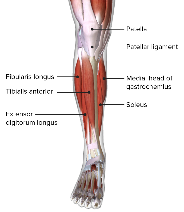
Leg Anatomy Muscles Ligaments And Tendons Leg muscles. your legs include many strong muscles. they allow you to do big and small movements. they also support your weight and stabilize your body so you can stand up straight. the muscles in your upper leg include your quadriceps and hamstrings. your calf muscles work with other muscles of the lower leg to help you move your feet. Anatomically, the leg is defined as the region of the lower limb below the knee. it consists of a posterior, anterior and lateral compartment. in accordance, the muscles of the leg are organized into three groups: anterior (dorsiflexor) group, which contains the tibialis anterior, extensor digitorum longus, fibularis tertius and extensor.

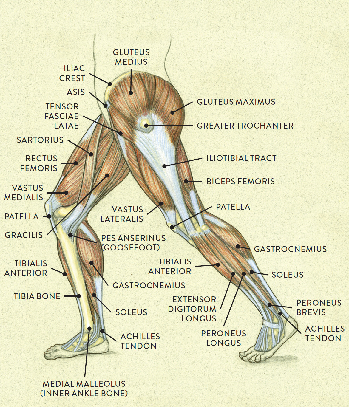
Leg Tendons And Ligaments Diagram Plantaris. this is a small muscle in the back of the lower leg. like the gastrocnemius and soleus, it’s involved in plantar flexion. tibialis muscles. these muscles are found on the front and. Gastrocnemius (calf muscle): one of the large muscles of the leg, it connects to the heel. it flexes and extends the foot, ankle, and knee. soleus: this muscle extends from the back of the knee to. It has an upper extremity, a shaft, and a lower extremity, all of which are full of various structural landmarks. several muscles attach to, and act on, the femur. they take full advantage of the mobility provided by two joints. the muscles of the thigh can be divided into three groups: anterior, medial, and posterior. The main parts of the knee joint are the femur, tibia, patella, and supporting ligaments. the condyles of the femur and of the tibia come in close proximity to form the main structure of the joint. the patella, commonly known as the ‘kneecap’, is a sesamoid bone that sits within the tendon of the quadriceps femoris.

Leg Tendons And Ligaments Diagram It has an upper extremity, a shaft, and a lower extremity, all of which are full of various structural landmarks. several muscles attach to, and act on, the femur. they take full advantage of the mobility provided by two joints. the muscles of the thigh can be divided into three groups: anterior, medial, and posterior. The main parts of the knee joint are the femur, tibia, patella, and supporting ligaments. the condyles of the femur and of the tibia come in close proximity to form the main structure of the joint. the patella, commonly known as the ‘kneecap’, is a sesamoid bone that sits within the tendon of the quadriceps femoris. The lower leg refers to the portion of the lower extremity between the knee and ankle. this area consists of bones, muscles, tendons, and nerves that all work together to allow the leg to function. Knee anatomy involves more than just muscles and bones. ligaments, tendons, and cartilage work together to connect the thigh bone, shin bone, and knee cap and allow the leg to bend back and forth like a hinge. the largest joint in the body, the knee is also one of the most easily injured. problems with any part of the knee's anatomy can result.

Leg Anatomy Concise Medical Knowledge The lower leg refers to the portion of the lower extremity between the knee and ankle. this area consists of bones, muscles, tendons, and nerves that all work together to allow the leg to function. Knee anatomy involves more than just muscles and bones. ligaments, tendons, and cartilage work together to connect the thigh bone, shin bone, and knee cap and allow the leg to bend back and forth like a hinge. the largest joint in the body, the knee is also one of the most easily injured. problems with any part of the knee's anatomy can result.

Leg Tendons Diagram

Comments are closed.