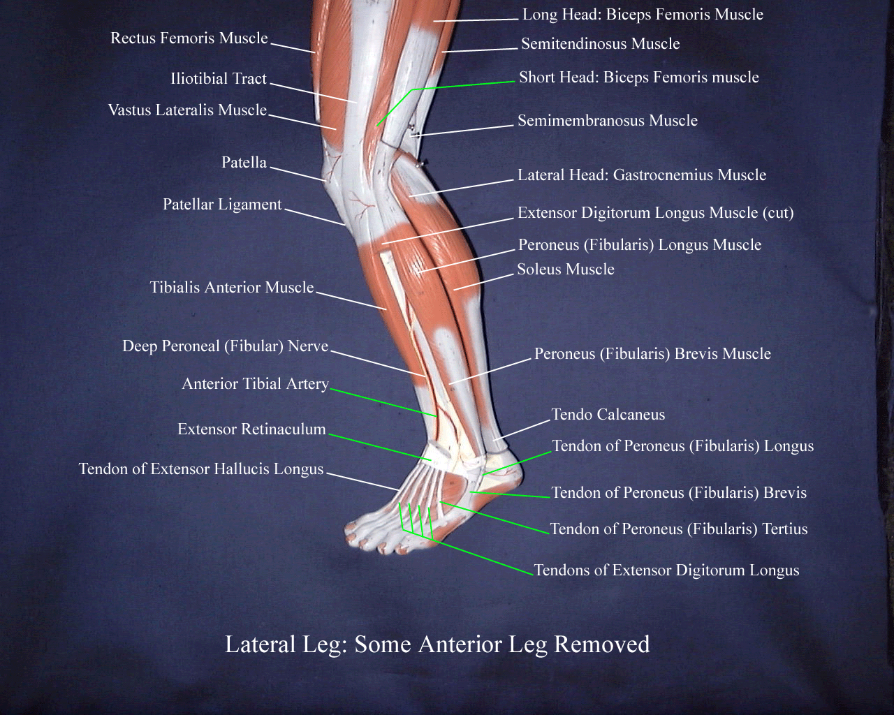
Laterallegmodeldeep

Laterallegmodeldeep Laterallegmodeldeep east tennessee state university © forsman, 2001. Take the quiz. there are two muscles in the lateral compartment of the leg; the fibularis longus and brevis (also known as peroneal longus and brevis). the common function of the muscles is eversion – turning the sole of the foot outwards. they are both innervated by the superficial fibular nerve.

Usa Lower Extremity Model Lower Leg Deep Lateral View Diagram Quizlet Leg muscles (musculi cruris) anatomically, the leg is defined as the region of the lower limb below the knee. it consists of a posterior, anterior and lateral compartment. in accordance, the muscles of the leg are organized into three groups: anterior (dorsiflexor) group, which contains the tibialis anterior, extensor digitorum longus. Explore the anatomy of leg muscles, including anterior and lateral regions, with teachmeanatomy's comprehensive guide. It has an upper extremity, a shaft, and a lower extremity, all of which are full of various structural landmarks. several muscles attach to, and act on, the femur. they take full advantage of the mobility provided by two joints. the muscles of the thigh can be divided into three groups: anterior, medial, and posterior. The main parts of the knee joint are the femur, tibia, patella, and supporting ligaments. the condyles of the femur and of the tibia come in close proximity to form the main structure of the joint. the patella, commonly known as the ‘kneecap’, is a sesamoid bone that sits within the tendon of the quadriceps femoris.

Laterallegmodel It has an upper extremity, a shaft, and a lower extremity, all of which are full of various structural landmarks. several muscles attach to, and act on, the femur. they take full advantage of the mobility provided by two joints. the muscles of the thigh can be divided into three groups: anterior, medial, and posterior. The main parts of the knee joint are the femur, tibia, patella, and supporting ligaments. the condyles of the femur and of the tibia come in close proximity to form the main structure of the joint. the patella, commonly known as the ‘kneecap’, is a sesamoid bone that sits within the tendon of the quadriceps femoris. Containing the fibularis longus muscle (flm) and fibularis brevis muscle (fbm), common fibular and superficial fibular nerves, and branches of the anterior tibial and fibular arteries, the lateral leg compartment is one of the four compartments of the leg.[1][2] structures with the term “peroneal” have been replaced with “fibular” for anatomical accuracy. the primary function of the. The flexor digitorum longus is a thin muscle and is located medially within the posterior leg. attachments: originates from the medial surface of the tibia and attaches to the plantar surfaces of the lateral four digits. actions: flexion of the lateral four toes. innervation: tibial nerve.

Deep Lateral View Of The Lower Limb Diagram Quizlet Containing the fibularis longus muscle (flm) and fibularis brevis muscle (fbm), common fibular and superficial fibular nerves, and branches of the anterior tibial and fibular arteries, the lateral leg compartment is one of the four compartments of the leg.[1][2] structures with the term “peroneal” have been replaced with “fibular” for anatomical accuracy. the primary function of the. The flexor digitorum longus is a thin muscle and is located medially within the posterior leg. attachments: originates from the medial surface of the tibia and attaches to the plantar surfaces of the lateral four digits. actions: flexion of the lateral four toes. innervation: tibial nerve.

Comments are closed.