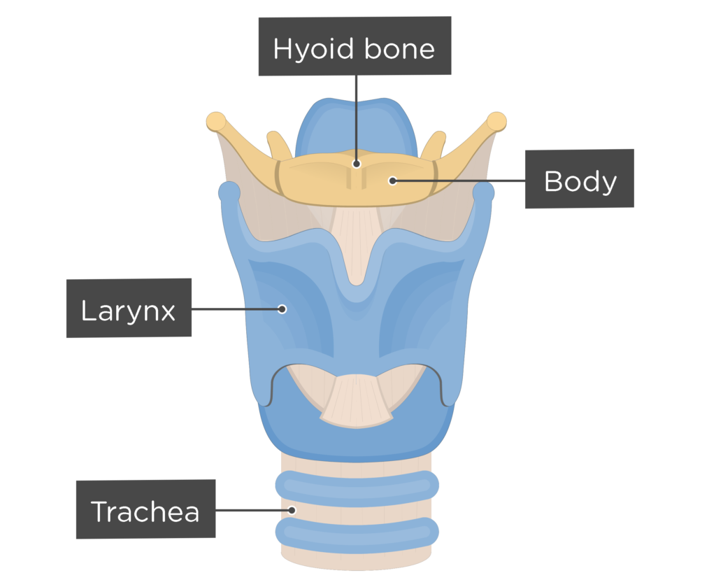
Hyoid Bone Labeled

Hyoid Bone Description Anatomy Function Britannica Learn about the hyoid bone, a u shaped bone in the anterior neck that is not attached to other bones. see its gross anatomy, muscular attachments, embryology and clinical significance. Learn about the hyoid bone, the only floating bone in your body that supports your tongue and helps you speak and swallow. find out how to locate, identify and care for your hyoid bone and its possible conditions.
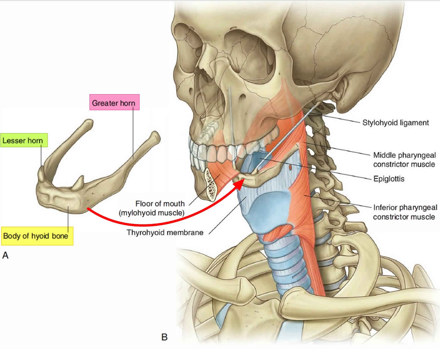
Hyoid Bone Anatomy Location Dislocation Fracture Hyoid Bone Syndrome The hyoid bone is a horseshoe shaped bone in the neck that is not attached to any other bones. it has a body and two pairs of horns, and provides attachment to muscles of the tongue, larynx and pharynx. Learn about the anatomy and function of the hyoid bone, a 'u' shaped bone in the anterior neck. see diagrams, 3d model and quiz to test your knowledge. The anatomy of the hyoid bone. the hyoid bone is a small horseshoe shaped bone located in the front of your neck. it sits between the chin and the thyroid cartilage and is instrumental in the function of swallowing and tongue movements. the little talked about hyoid bone is a unique part of the human skeleton for a number of reasons. Learn about the hyoid bone, a small u shaped bone in the neck that does not articulate with any other bone. see its anatomy, functions, attachments, and labeled diagram with parts and landmarks.
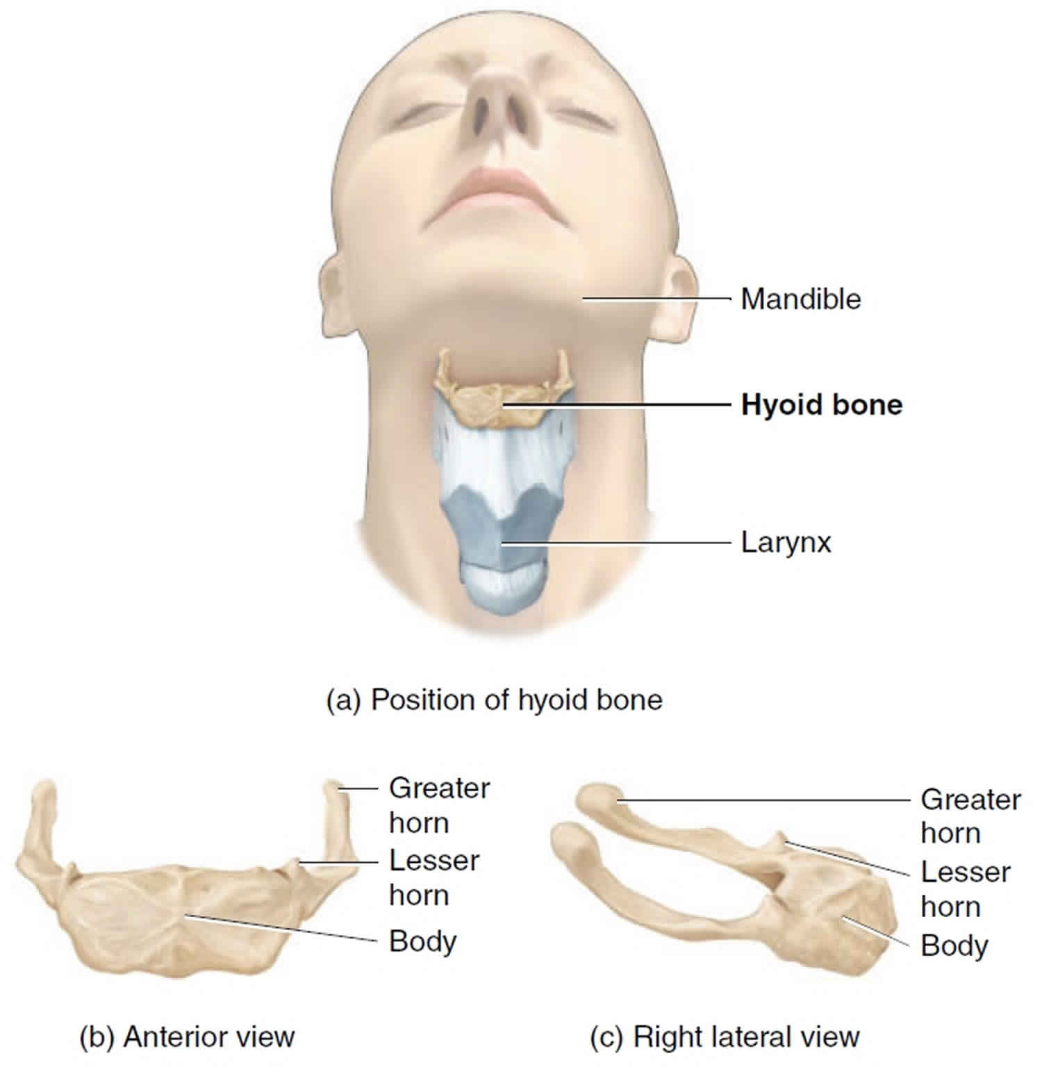
álbumes 93 Imagen De Fondo Donde Se Encuentra El Hueso Hioides El último The anatomy of the hyoid bone. the hyoid bone is a small horseshoe shaped bone located in the front of your neck. it sits between the chin and the thyroid cartilage and is instrumental in the function of swallowing and tongue movements. the little talked about hyoid bone is a unique part of the human skeleton for a number of reasons. Learn about the hyoid bone, a small u shaped bone in the neck that does not articulate with any other bone. see its anatomy, functions, attachments, and labeled diagram with parts and landmarks. Sternohyoid muscle: the muscle gets inserted in the inferior surface of the vocal bone. 2. omohyoid muscle: it inserts on the inferolateral surface of the vocal bone. 3. sternothyroid muscle: this muscle does not directly attach to the vocal bone. it inserts on the oblique line of the thyroid cartilage. 4. The hyoid bone (hyoid) is a small u shaped (horseshoe shaped) solitary bone situated in the midline of the neck anteriorly at the base of the mandible and posteriorly at the fourth cervical vertebra. its anatomical position is just superior to the thyroid cartilage. it is closely linked with an extended tendon muscular complex but not specifically interconnected to any adjacent bones and.
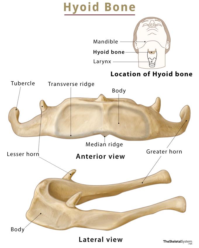
Hyoid Bone Location Functions Anatomy Labeled Diagram Sternohyoid muscle: the muscle gets inserted in the inferior surface of the vocal bone. 2. omohyoid muscle: it inserts on the inferolateral surface of the vocal bone. 3. sternothyroid muscle: this muscle does not directly attach to the vocal bone. it inserts on the oblique line of the thyroid cartilage. 4. The hyoid bone (hyoid) is a small u shaped (horseshoe shaped) solitary bone situated in the midline of the neck anteriorly at the base of the mandible and posteriorly at the fourth cervical vertebra. its anatomical position is just superior to the thyroid cartilage. it is closely linked with an extended tendon muscular complex but not specifically interconnected to any adjacent bones and.
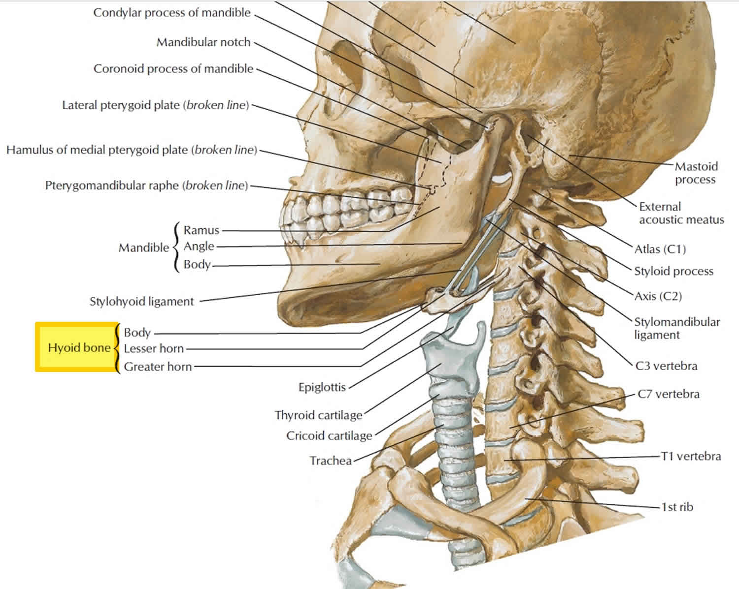
Hyoid Bone Anatomy Location Dislocation Fracture Hyoid Bone Syndrome
Human Hyoid Bone

Comments are closed.