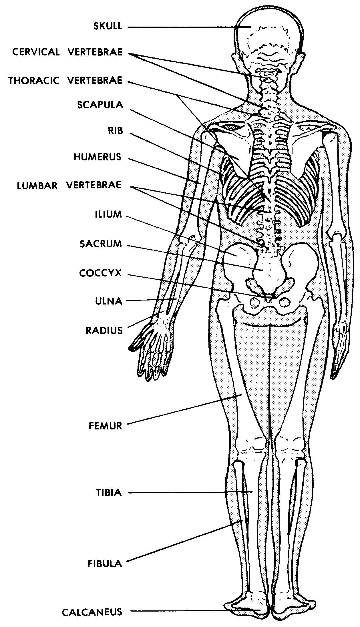
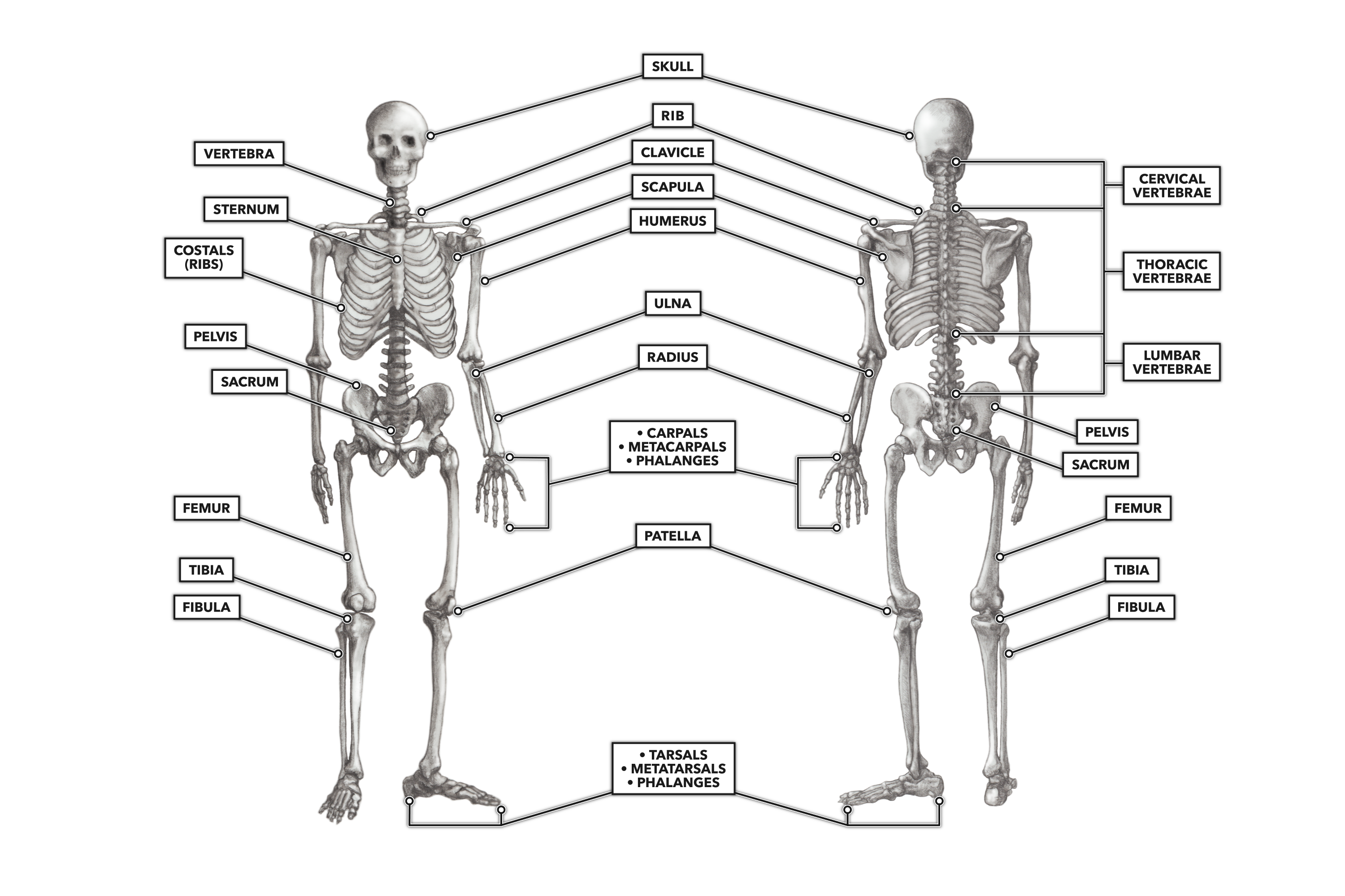
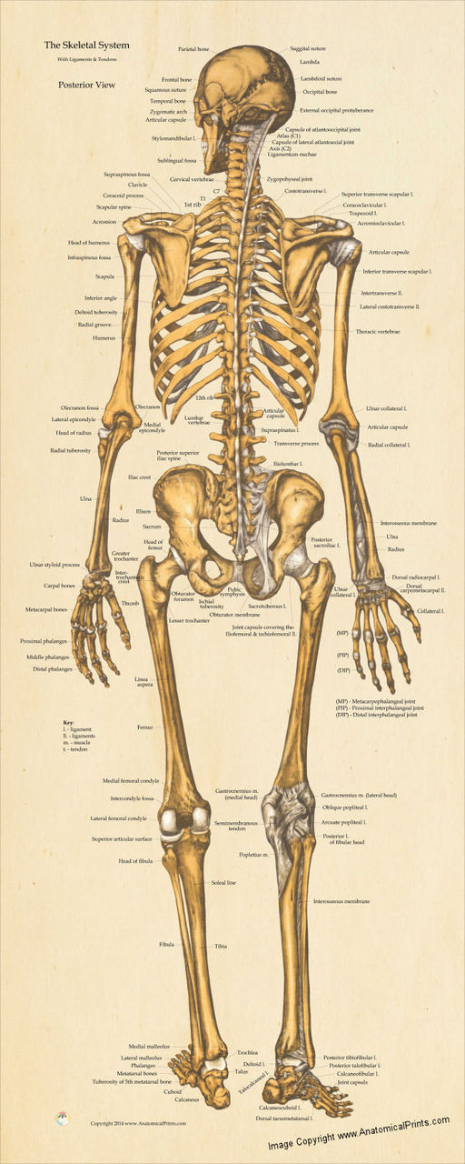
Human Skeleton Diagram Posterior View

Images 04 Skeletal System Basic Human Anatomy The skeletal system. explore the skeletal system with our interactive 3d anatomy models. learn about the bones, joints, and skeletal anatomy of the human body. the skeletal system includes all of the bones and joints in the body. each bone is a complex living organ that is made up of many cells, protein fibers, and minerals. Anatomy systems. skeletal system the skeletal system includes all of the bones and joints in the body. muscular system the muscular system is responsible for the movement of the human body. cardiovascular system the cardiovascular system consists of the heart, blood vessels, and the approximately 5 liters of blood that the blood vessels transport.

Human Skeleton Diagram Posterior View Human skeleton, the internal skeleton that serves as a framework for the body. this framework consists of many individual bones and cartilages. there also are bands of fibrous connective tissue —the ligaments and the tendons —in intimate relationship with the parts of the skeleton. this article is concerned primarily with the gross. Function. conditions. diagnosis. skeletal system photos and labeled diagrams can help you to understand the parts of the skeleton, how it's organized, the conditions that can affect it, and tests used to diagnose skeletal health. the skeletal system comprises 206 bones and has two main parts: the axial skeleton and the appendicular skeleton. Here’s a skeletal system diagram providing you with a broad overview of the two skeletons and the bones in the body: [main bones of the skeletal system (anterior view)] the axial skeleton is essentially the midline, or central core region, and consists of the bones of the skull (cranium) together with the bones of the trunk. Occipital bone. flat skull bone articulating with the parietal bone and atlas (first cervical vertebra), among others; it makes up the largest portion of the base of the skull. lateral view of skull.

Skeletal System Posterior View Poster Clinical Charts And Supplies Here’s a skeletal system diagram providing you with a broad overview of the two skeletons and the bones in the body: [main bones of the skeletal system (anterior view)] the axial skeleton is essentially the midline, or central core region, and consists of the bones of the skull (cranium) together with the bones of the trunk. Occipital bone. flat skull bone articulating with the parietal bone and atlas (first cervical vertebra), among others; it makes up the largest portion of the base of the skull. lateral view of skull. The skeletal system provides our body with shape and stability, as well as the protection of internal organs. it is composed of 206 bones that connect to each other via joints. accessory structures that support the skeletal system are the cartilage, ligaments, bursae and muscle tendons. the bone is a calcified hard tissue that presents the main. These are the images used in the video lecture and study guide. figure 4 2. a “typical synovial joint:–diagrammatic. figure 4 4. a typical vertebra (superior and side views). figure 4 5. the human thorax with bones of the shoulder region. figure 4 6. the human skull (front and side views).

Comments are closed.