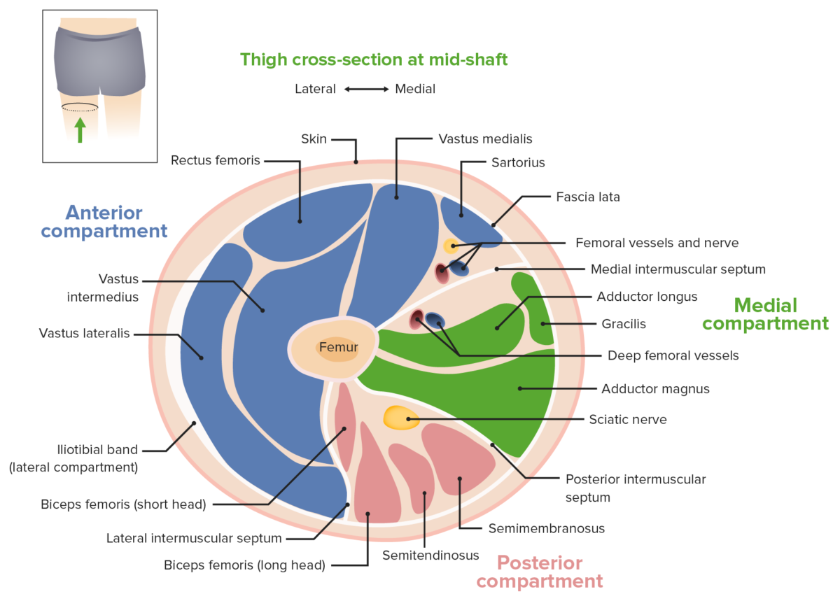
Gross Anatomy Glossary Cross Section Of The Mid Thigh Draw It To Know It

Gross Anatomy Glossary Cross Section Of The Mid Thigh Ditki Medical – rectus femoris and the vastus muscles are collectively referred to as the "quadriceps femoris." sartorius is anatomically categorized as an anterior compartment muscle due to its origin on the pelvis and its action on the thigh, but, it crosses obliquely over the quadriceps femoris and is visible within the medial compartment at the level of our cross section. 14. femur. 15. short head of biceps femoris. 16. sciatic nerve. 17. label muscular structures in a cross section of the leg learn with flashcards, games, and more — for free.

Gross Anatomy Glossary Medial Thigh Muscles Draw It To Know It Description: cross section through middle of thigh. a cross section of a male thigh can be seen. english labels. retrieved from website clinical anatomy of the university of british columbia. anatomical structures in item: musculus rectus femoris. musculus vastus medialis. musculus sartorius. Cross section of the thigh learn with flashcards, games, and more — for free. anatomy of teeth . 14 terms. flahertyhaley6. preview. endocrine system. 10 terms. Cross sections are two dimensional, axial views of gross anatomical structures seen in transverse planes. they are obtained by taking imaginary slices perpendicular to the main axis of organs, vessels, nerves, bones, soft tissue, or even the entire human body. cross sections provide the perception of ‘depth’, creating three dimensional. Read chapter 36 of the big picture: gross anatomy, medical course & step 1 review, 2nd edition online now, exclusively on accessmedicine. accessmedicine is a subscription based resource from mcgraw hill that features trusted medical content from the best minds in medicine.

Thigh Anatomy Concise Medical Knowledge Cross sections are two dimensional, axial views of gross anatomical structures seen in transverse planes. they are obtained by taking imaginary slices perpendicular to the main axis of organs, vessels, nerves, bones, soft tissue, or even the entire human body. cross sections provide the perception of ‘depth’, creating three dimensional. Read chapter 36 of the big picture: gross anatomy, medical course & step 1 review, 2nd edition online now, exclusively on accessmedicine. accessmedicine is a subscription based resource from mcgraw hill that features trusted medical content from the best minds in medicine. Three compartments of the leg: • anterior • posterior • lateral bones of the leg & footmembranesanterior septum • extends anterolaterally from the fibula and separates the anterior and lateral compartments. posterior septum extends posterolat. Big picture. the bone between the hip and the knee is the femur. it is the longest and strongest bone in the body. the femur articulates proximally with the acetabulum and distally with the tibia and patella. the knee joint is formed by articulations of the femur, tibia, and patella. the knee joint enables flexion, extension, and minimal.

Comments are closed.