Frontiers Image Based Differentiation Of Benign And Malignant

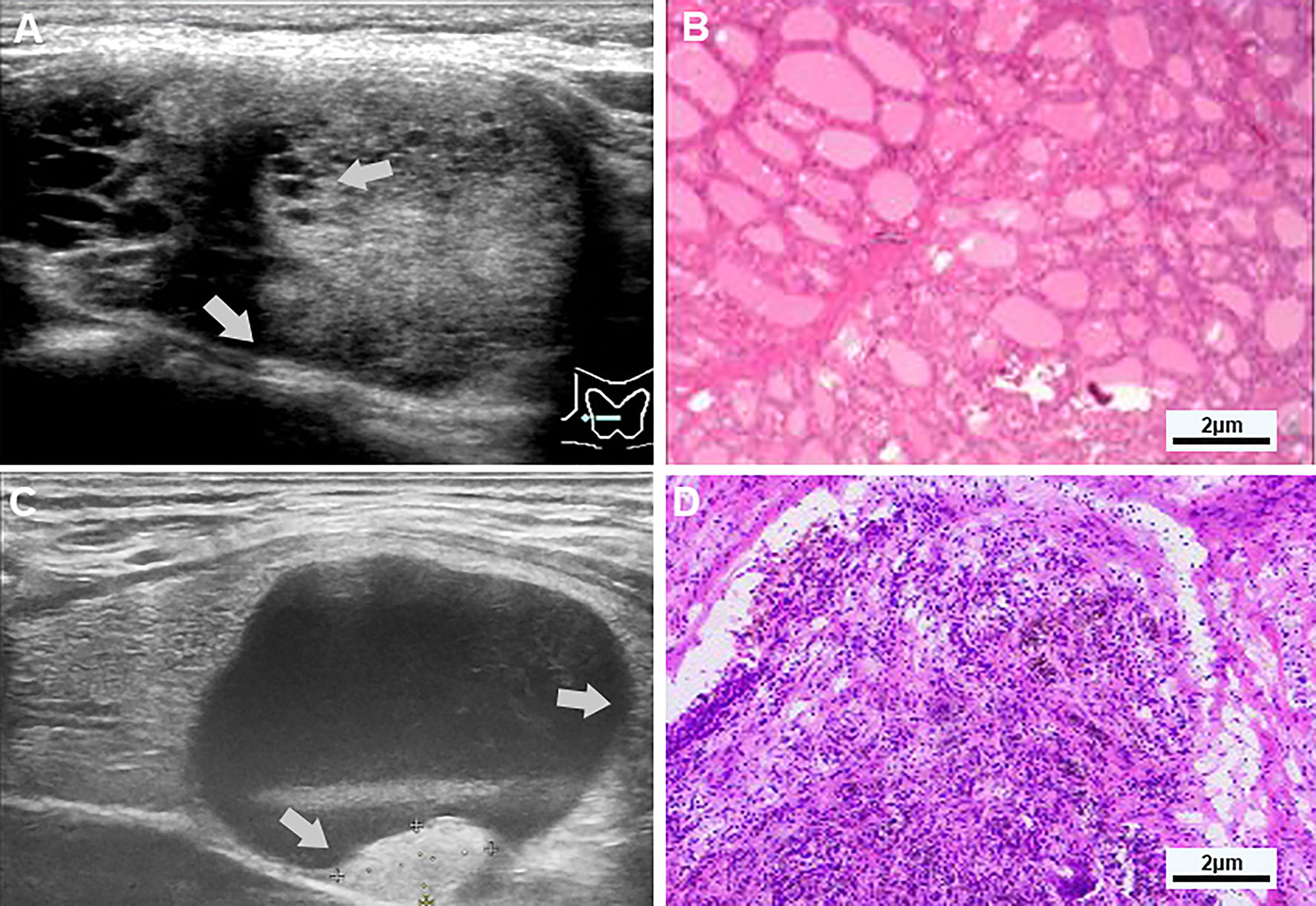
Frontiers Analysis Of Differential Diagnosis Of Benign And Malignant Unlike biopsy, these image based methods are noninvasive and more suitable screening tools. one of the main functions of medical imaging methods is to distinguish benign lesions from malignant tumors. a report suggested that ultrasound based differentiation of malignant and benign thyroid nodules has promising potential for clinical use . Non invasive image based diagnostic methods such as ct and mri are often considered essential screening tools for multiple types of tumors. for nf1 patients' lifelong regular follow ups, these radiological methods are currently used for tumor evaluation. however, no consensus was established on screening the malignant transformation of benign.

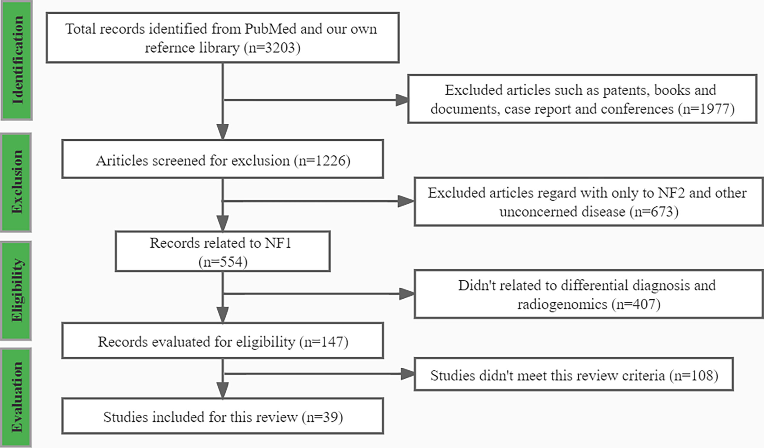
Frontiers Image Based Differentiation Of Benign And Malignant Radiology image based differentiation of benign and malignant tumors in nf1. this review comprehensively summarizes different image based methods used in distinguishing benign from malignant nf1 tumors, including ct, mri, pet ct, and ultrasound. this review also discusses the combination of radiology images and multiple omics disciplines, such. Citation: wang y, gao j, yin z, wen y, sun m and han r (2024) differentiation of benign and malignant parotid gland tumors based on the fusion of radiomics and deep learning features on ultrasound images. front. oncol. 14:1384105. doi: 10.3389 fonc.2024.1384105. received: 08 february 2024; accepted: 29 april 2024; published: 13 may 2024. The use of color duplex doppler and contrast agent adds valuable information for the differentiation of benign and malignant tumors.conclusion high resolution ultrasound is a well established, non. This review summarizes current studies of different imaging methods for differentiating benign and malignant tumors in nf1 and discussed the prospects of the usage of new tools such as radiogenomics and pet mri to distinguish mpnst from benign pnsts more precisely. neurofibromatosis type 1 (nf1) is a dominant hereditary disease characterized by the mutation of the nf1 gene, affecting 1 3000.

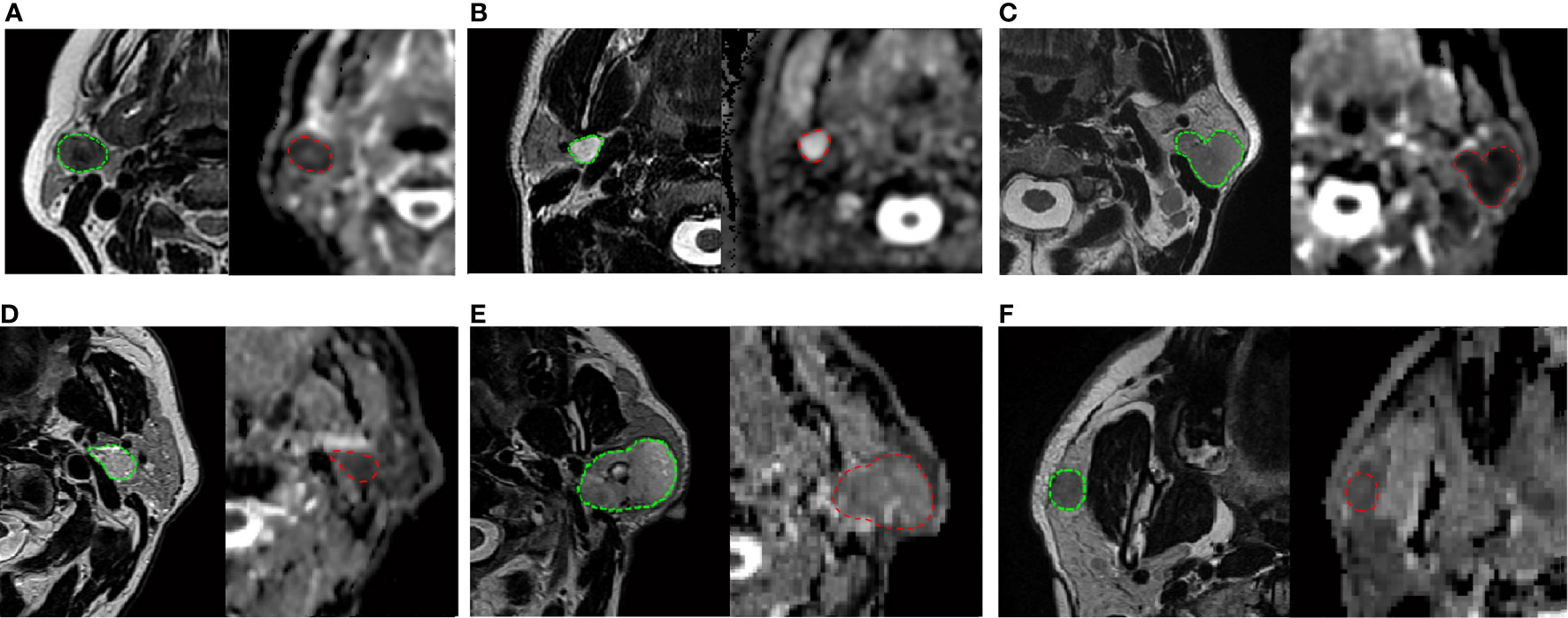
Frontiers Mri Based Radiomics To Differentiate Between Benign And The use of color duplex doppler and contrast agent adds valuable information for the differentiation of benign and malignant tumors.conclusion high resolution ultrasound is a well established, non. This review summarizes current studies of different imaging methods for differentiating benign and malignant tumors in nf1 and discussed the prospects of the usage of new tools such as radiogenomics and pet mri to distinguish mpnst from benign pnsts more precisely. neurofibromatosis type 1 (nf1) is a dominant hereditary disease characterized by the mutation of the nf1 gene, affecting 1 3000. Europe pmc is an archive of life sciences journal literature. do data resources managed by embl ebi and our collaborators make a difference to your work? if so, please take 10 minutes to fill in our survey, and help us make the case for why sustaining open data resources is critical for life sciences research. A total of 3791 parotid gland region images were cropped from the mr images. a label (pleomorphic adenoma and warthin tumor, malignant tumor or free of tumor), which was based on histology results, was assigned to each image. to train the deep learning model, these data were randomly divided into a training dataset (90%, comprising 3035 mr.

Comments are closed.