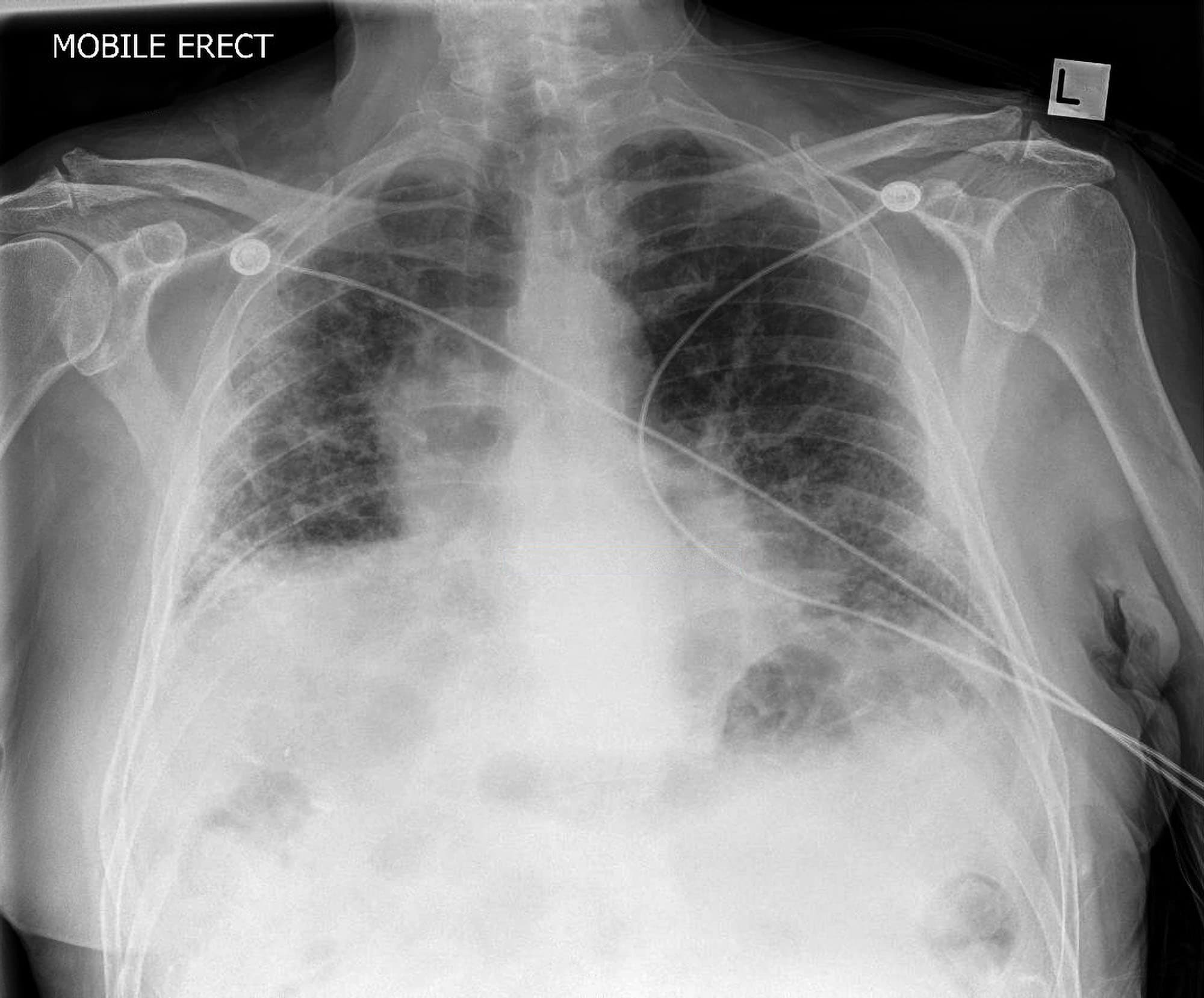
For The Love Of Med School Imaging Interstitial Lung Disease

For The Love Of Med School Imaging Interstitial Lung Disease Imaging interstitial lung disease. cxr patterns of interstitial lung disease ddx. peripheral reticular netlike pattern. (1) ipf. (2) connective tissue disease (ra, scleroderma, mctd) (3) pneumoconiosis (asbestosis) linear kerly a, b lines. (1) chf. Abstract. until today, computed tomography (ct) is the most important and valuable radiological modality to detect, analyze, and diagnose diffuse interstitial lung diseases (dild), based on the unsurpassed morphological detail provided by high resolution ct technique. in the past decade, there has been a shift from an isolated histopathological.

For The Love Of Med School Imaging Interstitial Lung Disease The montage of glyphs for ct scans of 372 ltrc subjects demonstrates the spectrum of parenchymal abnormalities. subjects in the ltrc shown here represent a variety of ild, copd, and mixed parenchymal and or airway diseases including uip, nonspecific interstitial pneumonitis, hypersensitivity pneumonitis, emphysema, bronchiolitis, and combined pulmonary fibrosis and emphysema. The clinical importance of interstitial lung abnormality (ila) is increasingly recognized. in july 2020, the fleischner society published a position paper about ila. the purposes of this article are to summarize the definition, existing evidence, clinical management, and unresolved issues for ila from a radiologic standpoint and to provide a practical guide for radiologists. ila is a common. Magnetic resonance imaging (mri) has been emerging as an imaging modality to assess interstitial lung diseases (ild). an optimal chest mri protocol for ilds should include non contrast breath holding sequences, steady state free precession sequences, and contrast enhanced sequences. one of the main mri applications in ilds is the differentiation between areas of active inflammation (i.e. Interstitial lung disease (ild) is an umbrella term that encompasses a large number of disorders that are characterized by diffuse cellular infiltrates in a periacinar location. the spectrum of conditions included is broad, ranging from occasional self limited inflammatory processes to severe debilitating fibrosis of the lungs. terminology.

For The Love Of Med School Imaging Interstitial Lung Disease Magnetic resonance imaging (mri) has been emerging as an imaging modality to assess interstitial lung diseases (ild). an optimal chest mri protocol for ilds should include non contrast breath holding sequences, steady state free precession sequences, and contrast enhanced sequences. one of the main mri applications in ilds is the differentiation between areas of active inflammation (i.e. Interstitial lung disease (ild) is an umbrella term that encompasses a large number of disorders that are characterized by diffuse cellular infiltrates in a periacinar location. the spectrum of conditions included is broad, ranging from occasional self limited inflammatory processes to severe debilitating fibrosis of the lungs. terminology. Patients with interstitial lung disease often present with breathlessness, chronic cough, inspiratory crackles on auscultation, and abnormal spirometry.1 2 over 200 different types of disease can cause thickening of the pulmonary interstitium, with the common final pathway for many of these being fibrosis (figs 1 and 2)⇓ ⇓.3 4 some forms of interstitial lung disease can be life threatening. Analogous to the finding of coronary artery calcifications as a harbinger of early heart disease, interstitial lung abnormality (ila) is often an early finding of pulmonary fibrosis. ila may progress to the commonly recognized ct features of idiopathic pulmonary fibrosis (ipf) and is correlated with worsened clinical outcomes.

For The Love Of Med School Imaging Interstitial Lung Disease Patients with interstitial lung disease often present with breathlessness, chronic cough, inspiratory crackles on auscultation, and abnormal spirometry.1 2 over 200 different types of disease can cause thickening of the pulmonary interstitium, with the common final pathway for many of these being fibrosis (figs 1 and 2)⇓ ⇓.3 4 some forms of interstitial lung disease can be life threatening. Analogous to the finding of coronary artery calcifications as a harbinger of early heart disease, interstitial lung abnormality (ila) is often an early finding of pulmonary fibrosis. ila may progress to the commonly recognized ct features of idiopathic pulmonary fibrosis (ipf) and is correlated with worsened clinical outcomes.

For The Love Of Med School Imaging Interstitial Lung Disease

Interstitial Lung Disease Chest X Ray Medschool

Comments are closed.