Figure 2 From Reconstruction Of The Nasal Septum Using Perforated And

Figure 2 From Reconstruction Of The Nasal Septum Using Perforated And Results: fifty patients underwent septal reconstruction using unperforated (first 26 patients) or perforated (next 24 patients) polydioxanone foil. median total postoperative follow up was 51.5 months (range, 34 60 months) for unperforated foil and 20.5 months (range, 12 31 months) for perforated foil. all the patients were reviewed for. Temporoparietal fascia grafts have a proven success rate in the repair of anterior skull base defects 12 and have been used with success for other reconstructive procedures, including pharyngeal repair 13 and microtia reconstruction. 14 two prior retrospective case series have evaluated the combined use of pds plates with a tpf graft for nasal.

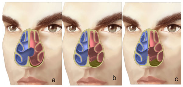
Figure 2 From Repair Of Nasal Septal Perforation Using A Simple Figure 2. extracorporeal septoplasty using bilateral polydioxanone foil battens. a deficient dorsum is reconstructed by transposing a strip of posterior septal cartilage stabilized between 2 layers of polydioxanone (a and b), before reinserting into the nose (c). "reconstruction of the nasal septum using perforated and unperforated polydioxanone foil.". Objective to present our experience of reconstruction of the nasal septum using perforated and unperforated foils, particularly with respect to functional and aesthetic sequelae. methods a retrospective medical record review of a prospectively conducted case series was undertaken of all consecutive patients who underwent septal reconstruction using polydioxanone foil in a 4 year period. Separated inferior turbinate flap with 1 to 2 cm attachment to the lateral nasal wall. then a pds plate is cut to size approximately 1 cm larger than the nasal septal perforation. a piece of adm (0.33–0.76 mm thick) is secured to the pds plate using a 5–0 pds suture. this pds plate adm complex is implanted between the nasal septal flaps. Figure 2: perpendicular plate of ethmoid attaches to cribriform plate vascular supply (figures 3 5) an understanding of the vascular supply is important if large mucoperiosteal flaps are to be used to repair a septal perforation (figure 3). venous outflow generally fol lows the arterial blood supply. the nasal septum has both internal (ica).

Septal Perforation Rhinoplasty Archive Separated inferior turbinate flap with 1 to 2 cm attachment to the lateral nasal wall. then a pds plate is cut to size approximately 1 cm larger than the nasal septal perforation. a piece of adm (0.33–0.76 mm thick) is secured to the pds plate using a 5–0 pds suture. this pds plate adm complex is implanted between the nasal septal flaps. Figure 2: perpendicular plate of ethmoid attaches to cribriform plate vascular supply (figures 3 5) an understanding of the vascular supply is important if large mucoperiosteal flaps are to be used to repair a septal perforation (figure 3). venous outflow generally fol lows the arterial blood supply. the nasal septum has both internal (ica). After complete tunnelling between forehead and nasal dorsum the flap will be rotated 180° through the open nasal roof into the separated mucosal margins of the perforation (figure 9 a) figure 9. closure of a septum perforation using the subcutanous galea periost flap. a: preoperative perforation. b: during surgery with sutures positioned. c: 6. The most common causes of nasal septal perforations are trauma, iatrogenic e.g. septoplasty, and pharmacologic e.g. intranasal corticosteroids and cocaine abuse. other etiologies such as vasculitis, malignancy and infection should be considered when investigating a patient when a cause is not apparent. most septal perforations go unnoticed.

Pdf Nasal Septal Perforation Repair Using An Inferior Turbinate Flap After complete tunnelling between forehead and nasal dorsum the flap will be rotated 180° through the open nasal roof into the separated mucosal margins of the perforation (figure 9 a) figure 9. closure of a septum perforation using the subcutanous galea periost flap. a: preoperative perforation. b: during surgery with sutures positioned. c: 6. The most common causes of nasal septal perforations are trauma, iatrogenic e.g. septoplasty, and pharmacologic e.g. intranasal corticosteroids and cocaine abuse. other etiologies such as vasculitis, malignancy and infection should be considered when investigating a patient when a cause is not apparent. most septal perforations go unnoticed.

Comments are closed.