Fetal Ventriculomegaly Ppt

Fetal Ventriculomegaly Ppt This document discusses ventriculomegaly (vm), which is the enlargement of the lateral cerebral ventricles. vm has many potential causes including infections, vascular issues, hydrocephalus, malformations, or genetic abnormalities. it can range from mild to severe. evaluation involves detailed ultrasound exams and may include fetal mri or. Ventriculomegaly is defined as dilation of the fetal cerebral ventricles and is a relatively common finding on prenatal ultrasound. the purpose of this document is to review the diagnosis, evaluation, and management of mild fetal ventriculomegaly. when enlargement of the lateral ventricles (≥10 mm) is identified, a thorough evaluation should be performed, including detailed sonographic.

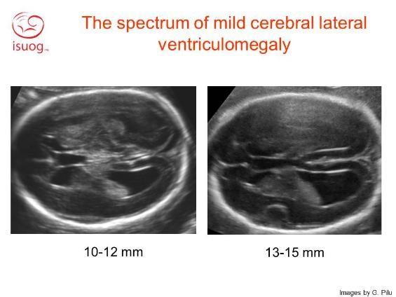
Fetal Ventriculomegaly Ppt Ventriculomegaly is the term used to describe cerebral ventricular dilation unrelated to increased cerebrospinal fluid (csf) pressure, such as dilation due to brain dysgenesis or atrophy. hydrocephalus is the term used to describe pathologic dilation of the brain's ventricular system due to increased csf pressure; obstruction is a common. Fetal cerebral ventriculomegaly is defined as an atrial diameter of ≥10 mm on prenatal ultrasound in the second and third trimesters of pregnancy.1–3 the terms hydrocephalus and ventriculomegaly are often used interchangeably. more often, however, the term hydrocephalus is used to describe pathologic dilation of the brain’s ventricular system because of increased cerebrospinal fluid (csf. Fetal cerebral ventriculomegaly is a relatively common finding, observed during approximately 1% of obstetric ultrasounds. in the second and third trimester, mild (≥10 mm) and severe ventriculomegaly (≥15 mm) are defined according to the measurement of distal lateral ventricles that is included in the routine sonographic examination of central nervous system. Fetal brain anomalies. fetal brain anomalies were discussed including ventriculomegaly, agenesis of the corpus callosum, and dandy walker malformations. ventriculomegaly is enlargement of the lateral ventricles and can be mild (10 15mm) or severe (>15mm). it requires ruling out associated anomalies, infections, and chromosomal abnormalities.

Fetal Ventriculomegaly Ppt Fetal cerebral ventriculomegaly is a relatively common finding, observed during approximately 1% of obstetric ultrasounds. in the second and third trimester, mild (≥10 mm) and severe ventriculomegaly (≥15 mm) are defined according to the measurement of distal lateral ventricles that is included in the routine sonographic examination of central nervous system. Fetal brain anomalies. fetal brain anomalies were discussed including ventriculomegaly, agenesis of the corpus callosum, and dandy walker malformations. ventriculomegaly is enlargement of the lateral ventricles and can be mild (10 15mm) or severe (>15mm). it requires ruling out associated anomalies, infections, and chromosomal abnormalities. ÐÏ à¡± á> þÿ ü þÿÿÿþÿÿÿð ñ ò ó ô õ ö ÷ ø ù ú û. Fetal ventriculomegaly (vm) refers to the enlargement of the cerebral ventricles in utero. it is associated with the postnatal diagnosis of hydrocephalus. vm is clinically diagnosed on ultrasound and is defined as an atrial diameter greater than 10 mm. because of the anatomic detailed seen with advanced imaging, vm is often further characterized by fetal magnetic resonance imaging (mri). fetal.

Ventriculomegaly South West Fetal Network ÐÏ à¡± á> þÿ ü þÿÿÿþÿÿÿð ñ ò ó ô õ ö ÷ ø ù ú û. Fetal ventriculomegaly (vm) refers to the enlargement of the cerebral ventricles in utero. it is associated with the postnatal diagnosis of hydrocephalus. vm is clinically diagnosed on ultrasound and is defined as an atrial diameter greater than 10 mm. because of the anatomic detailed seen with advanced imaging, vm is often further characterized by fetal magnetic resonance imaging (mri). fetal.

Fetal Ventriculomegaly American Journal Of Obstetrics Gynecology

Comments are closed.