Fetal Ventriculomegaly Lurie Children S

Fetal Ventriculomegaly Lurie Children S Fetal ventriculomegaly. fetal ventriculomegaly is a congenital finding that affects the brain. the contents of the brain consist primarily of brain tissue, blood and cerebrospinal fluid (csf). enlargement of the ventricular system, the fluid filled spaces in the brain, can be caused by the overproduction of csf, inadequate brain development or. To learn more, please e mail the ann & robert h. lurie children’s hospital of chicago foundation at [email protected] or call 312.227.7500. the chicago institute for fetal health offers comprehensive pre birth counseling and pediatric care planning for pregnant women with fetal complications.

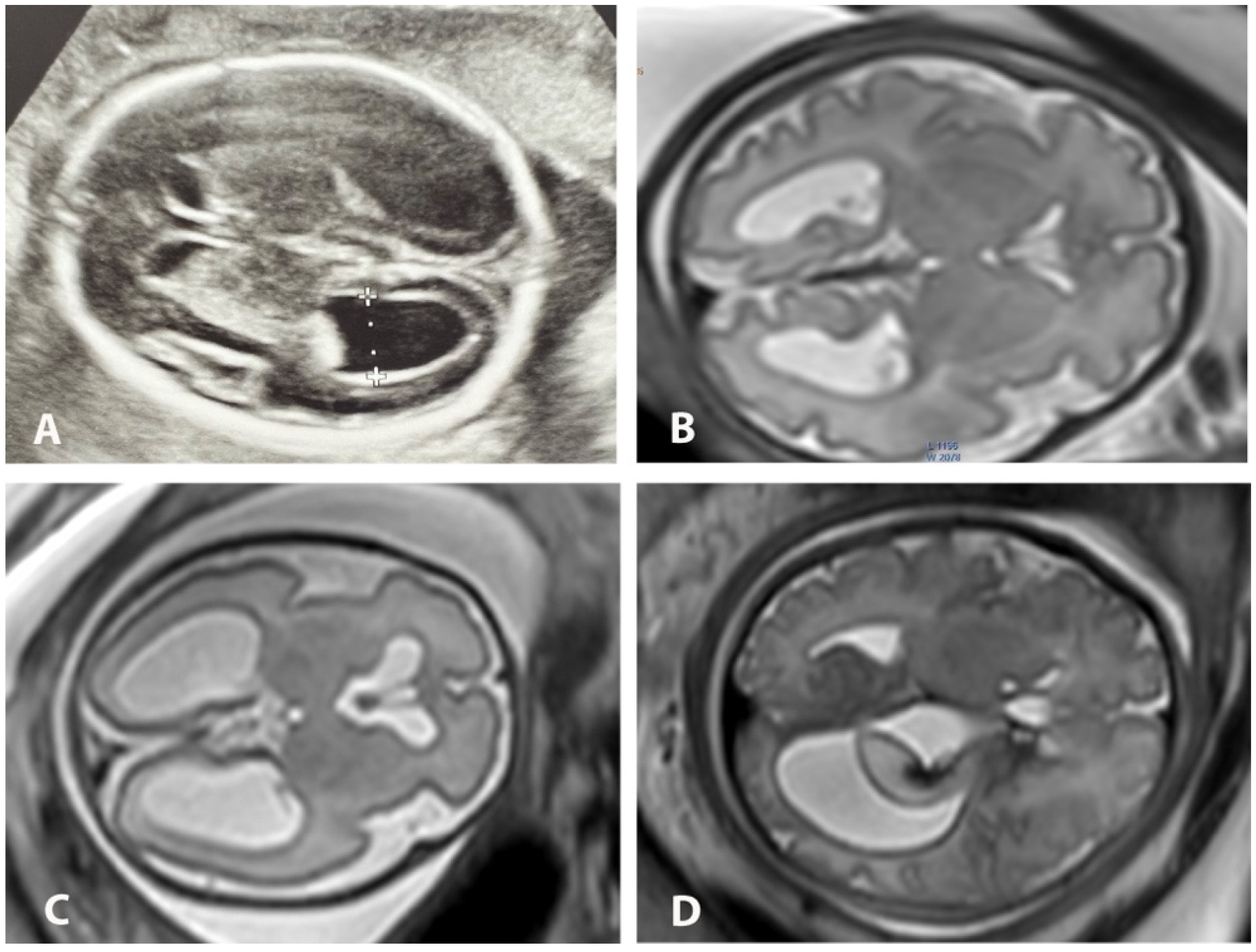
Ventriculomegaly Fetal Ultrasound Dr. robin bowman, lurie children's pediatric neurosurgeon, director of the multidisciplinary spina bifida center at lurie children’s and co director of fetal neurosurgery at the chicago institute for fetal health at lurie children’s, and along with lurie children's pediatric neurosurgeon and chief informatics officer dr. tord alden led the four part “fetal neurosurgery” section at the. Fetal cerebral ventriculomegaly, defined as dilation of cerebral ventricles at the level of the atria (≥10 mm), is one of the most common fetal neurological disorders identified prenatally by neuroimaging. 1 6 existing data suggest that cerebral ventriculomegaly affects up to 1% of pregnancies throughout gestation. 6 the presence of fetal ventriculomegaly can also alert clinicians to the. Ventriculomegaly is defined as dilation of the fetal cerebral ventricles and is a relatively common finding on prenatal ultrasound. the purpose of this document is to review the diagnosis, evaluation, and management of mild fetal ventriculomegaly. when enlargement of the lateral ventricles (≥10 mm) is identified, a thorough evaluation should be performed, including detailed sonographic. Fetal cerebral ventriculomegaly is one of the most common fetal neurological disorders identified prenatally by neuroimaging. the challenges in the evolving landscape of conditions like fetal cerebral ventriculomegaly involve accurate diagnosis and how best to provide prenatal counseling regarding prognosis as well as postnatal management and care of the infant. the purpose of this narrative.

Ventriculomegaly Fetal Ultrasound Ventriculomegaly is defined as dilation of the fetal cerebral ventricles and is a relatively common finding on prenatal ultrasound. the purpose of this document is to review the diagnosis, evaluation, and management of mild fetal ventriculomegaly. when enlargement of the lateral ventricles (≥10 mm) is identified, a thorough evaluation should be performed, including detailed sonographic. Fetal cerebral ventriculomegaly is one of the most common fetal neurological disorders identified prenatally by neuroimaging. the challenges in the evolving landscape of conditions like fetal cerebral ventriculomegaly involve accurate diagnosis and how best to provide prenatal counseling regarding prognosis as well as postnatal management and care of the infant. the purpose of this narrative. Ventriculomegaly is the term used to describe cerebral ventricular dilation unrelated to increased cerebrospinal fluid (csf) pressure, such as dilation due to brain dysgenesis or atrophy. hydrocephalus is the term used to describe pathologic dilation of the brain's ventricular system due to increased csf pressure; obstruction is a common. Fetal cerebral ventriculomegaly is defined as an atrial diameter of ≥10 mm on prenatal ultrasound in the second and third trimesters of pregnancy.1–3 the terms hydrocephalus and ventriculomegaly are often used interchangeably. more often, however, the term hydrocephalus is used to describe pathologic dilation of the brain’s ventricular system because of increased cerebrospinal fluid (csf.

Diagnostics Free Full Text Concordance Between Us And Mri Two Ventriculomegaly is the term used to describe cerebral ventricular dilation unrelated to increased cerebrospinal fluid (csf) pressure, such as dilation due to brain dysgenesis or atrophy. hydrocephalus is the term used to describe pathologic dilation of the brain's ventricular system due to increased csf pressure; obstruction is a common. Fetal cerebral ventriculomegaly is defined as an atrial diameter of ≥10 mm on prenatal ultrasound in the second and third trimesters of pregnancy.1–3 the terms hydrocephalus and ventriculomegaly are often used interchangeably. more often, however, the term hydrocephalus is used to describe pathologic dilation of the brain’s ventricular system because of increased cerebrospinal fluid (csf.

Fetal Ventriculomegaly American Journal Of Obstetrics Gynecology

Comments are closed.