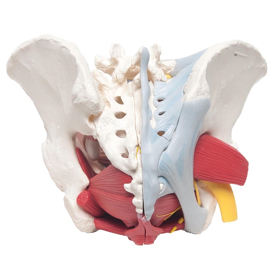
Female Pelvic Ligaments

Anatomical Models Of Female Pelvis With Ligaments Vessels Nerves The ligaments of the female reproductive tract are a series of structures that support the internal female genitalia in the pelvis. the ligaments of the female reproductive tract can be divided into three categories: broad ligament – a sheet of peritoneum, associated with both the uterus and ovaries. uterine ligaments – ligaments primarily. The pelvis contains a large number of organs, bones, muscles, and ligaments, so many conditions can affect the entire pelvis or parts within it. some conditions that can affect the female pelvis.

Standardized Terminology Of Apical Structures In The Female Pelvis The pelvis's bony integrity is supported by various ligaments that lend crucial flexible strength to the pelvic cavity and support some of the internal pelvic structures.[1] the principal ligaments are sacrotuberous, sacrospinous, and iliolumbar, and in the female pelvis, there are further ligaments to support the ovaries and uterus (see image. pelvic ligaments).[2] trauma or stretching of. The female reproductive system is made up of external and internal organs. the external organs lie in an area called the vulva, and they include the labia, the clitoris, and the vaginal opening. the internal reproductive organs can be found within the pelvic cavity, and they include the ovaries, which produce the female sex cells, called. The cardinal ligaments are attached to the side of the uterine cervix and the vaginal fornix, specifically its lateral parts. the cardinal ligaments are traversed by the ureters and pelvic blood vessels which can easily be injured during pelvic surgery. the cardinal ligaments are paired structures providing support to the female pelvic organs. Introduction. pelvic surgery requires a comprehensive knowledge of the pelvic anatomy to safely attain access, maximize exposure, ensure hemostasis, and avoid injury to viscera, blood vessels, and nerves. the anatomy of the female genital tract and lower urinary and gastrointestinal tracts relevant to the surgeon performing laparotomy or.

Surface Anatomy Female Pelvis The cardinal ligaments are attached to the side of the uterine cervix and the vaginal fornix, specifically its lateral parts. the cardinal ligaments are traversed by the ureters and pelvic blood vessels which can easily be injured during pelvic surgery. the cardinal ligaments are paired structures providing support to the female pelvic organs. Introduction. pelvic surgery requires a comprehensive knowledge of the pelvic anatomy to safely attain access, maximize exposure, ensure hemostasis, and avoid injury to viscera, blood vessels, and nerves. the anatomy of the female genital tract and lower urinary and gastrointestinal tracts relevant to the surgeon performing laparotomy or. The female reproductive tract is all located within the pelvis. it is made up of the vulva, the vagina, the cervix, the uterus, the fallopian tubes and the ovaries. these organs are supported in the pelvis by ligaments. the vulva refers to the external female genitalia. its anatomical structure can be broken down further into the mons pubis. Imagine the vagina and uterus forming a slightly tilted capital p in a sagittal section of the female pelvis. this organ is divided into three parts, the body, the isthmus and the cervix, which are supported by several ligaments and pelvic muscles. ligaments of the uterus. in this discussion, a "ligament" refers.

Comments are closed.