Chest Radiography Basics Of Interstitial Lung Disease

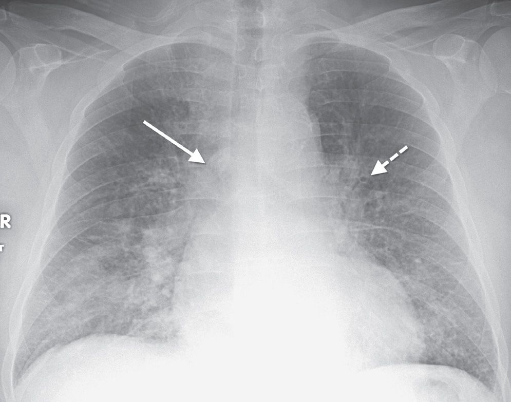
Interstitial Lung Disease Radiology Key Interstitial lung disease (ild) is an umbrella term that encompasses a large number of disorders that are characterized by diffuse cellular infiltrates in a periacinar location. the spectrum of conditions included is broad, ranging from occasional self limited inflammatory processes to severe debilitating fibrosis of the lungs. terminology. H eart failure. h istiocytosis (langerhans cell histiocytosis) ild, interstitial lung disease. ild may result in four patterns of abnormal opacity on chest radiographs and ct scans: linear, reticular, nodular, and reticulonodular (fig. 3.1). these patterns are more accurately and specifically defined on ct.

Hrct Chest In Interstitial Lung Disease Chest Radiography Basics Publicationdate 2007 12 20 update 2022 03 15. in this review we present the key findings in the most common interstitial lung diseases. there are numerous interstitial lung diseases, but in clinical practice only about ten diseases account for approximately 90% of cases. Diffuse interstitial lung diseases (dilds) comprise a huge number of diseases which diffusely involve the lung parenchyma. the dilds have been subcategorized into (a) dilds that have a known etiology, (b) the idiopathic interstitial pneumonias, (c) the granulomatous dilds, and (d) a group of diffuse lung diseases that include langerhans cell histiocytosis and lymphangioleiomyomatosis. hrct. The clinical importance of interstitial lung abnormality (ila) is increasingly recognized. in july 2020, the fleischner society published a position paper about ila. the purposes of this article are to summarize the definition, existing evidence, clinical management, and unresolved issues for ila from a radiologic standpoint and to provide a practical guide for radiologists. ila is a common. Abstract. until today, computed tomography (ct) is the most important and valuable radiological modality to detect, analyze, and diagnose diffuse interstitial lung diseases (dild), based on the unsurpassed morphological detail provided by high resolution ct technique. in the past decade, there has been a shift from an isolated histopathological.

Interstitial Lung Disease Uip Radiology At St Vincent S University The clinical importance of interstitial lung abnormality (ila) is increasingly recognized. in july 2020, the fleischner society published a position paper about ila. the purposes of this article are to summarize the definition, existing evidence, clinical management, and unresolved issues for ila from a radiologic standpoint and to provide a practical guide for radiologists. ila is a common. Abstract. until today, computed tomography (ct) is the most important and valuable radiological modality to detect, analyze, and diagnose diffuse interstitial lung diseases (dild), based on the unsurpassed morphological detail provided by high resolution ct technique. in the past decade, there has been a shift from an isolated histopathological. The diffuse parenchymal lung diseases (dpld), often collectively referred to as the interstitial lung diseases (ilds), are a heterogeneous group of disorders that are classified together because of similar clinical, radiographic, physiologic, or pathologic manifestations (algorithm 1) [1 5]. the descriptive term "interstitial" reflects the. Based on currently available, scientific evidence, however, your doctor may recommend: corticosteroid medications. many people diagnosed with interstitial lung diseases are initially treated with a corticosteroid (prednisone), sometimes in combination with other drugs that suppress the immune system.

Comments are closed.