Brain Ventricle Of A Baby

Brain Ventricles Anatomy Ultrasound can measure the fetal brain's ventricle size. these ultrasound measurements can be repeated during the rest of pregnancy to monitor the ventricle size. depending on the size and growth of the ventricles, your doctor may refer you to our prenatal pediatrics institute for further testing to evaluate your baby's condition. tests may. The severity of fetal ventriculomegaly ranges from mild to severe based on the measurement of the ventricle. in a typical fetal brain, the ventricles are less than 10 millimeters (mm) wide — or about the width of a pea. mild: the ventricles are between 10 mm to 12 mm. moderate: the ventricles are between 13 mm to 15 mm.

Fetal Brain At 12 Weeks Cerebral Ventricles 3d Ultrasound Youtube Certain infections, particularly those caused by the torch group of pathogens (toxoplasmosis, other agents, rubella, cytomegalovirus, and herpes simplex), can potentially affect the baby’s brain development and lead to enlarged ventricles. complications during birth can also sometimes result in enlarged ventricles. Ventriculomegaly is a condition in which the brain ventricles are enlarged due to build up of cerebrospinal fluid (csf), a fluid that protects the brain and spinal cord. the severity of ventriculomegaly depends on how enlarged the brain is. in some cases, fluid keeps building up, causing hydrocephalus. Enlargement of the ventricular system, the fluid filled spaces in the brain, can be caused by the overproduction of csf, inadequate brain development or destruction of brain tissue. in a normal fetal brain, the ventricles are less than 10 mm wide. when the ventricles are between 10 mm and 15 mm wide, the baby is diagnosed with mild. Ventriculomegaly is defined as dilation of the fetal cerebral ventricles and is a relatively common finding on prenatal ultrasound. the purpose of this document is to review the diagnosis, evaluation, and management of mild fetal ventriculomegaly. when enlargement of the lateral ventricles (≥10 mm) is identified, a thorough evaluation should be performed, including detailed sonographic.

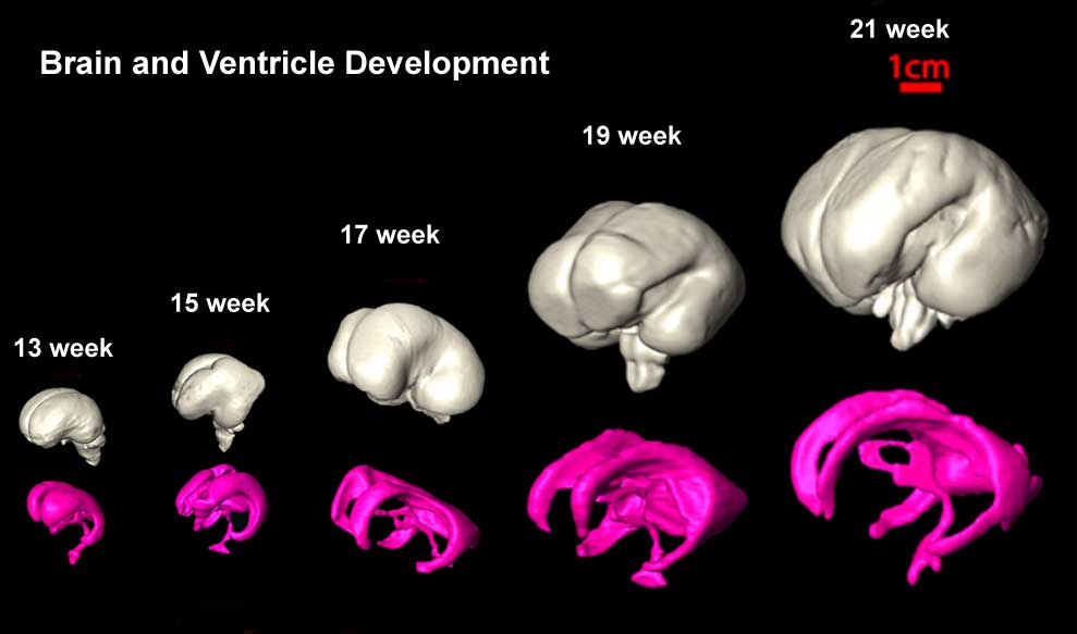
Brain Ventricle Of A Baby Youtube Enlargement of the ventricular system, the fluid filled spaces in the brain, can be caused by the overproduction of csf, inadequate brain development or destruction of brain tissue. in a normal fetal brain, the ventricles are less than 10 mm wide. when the ventricles are between 10 mm and 15 mm wide, the baby is diagnosed with mild. Ventriculomegaly is defined as dilation of the fetal cerebral ventricles and is a relatively common finding on prenatal ultrasound. the purpose of this document is to review the diagnosis, evaluation, and management of mild fetal ventriculomegaly. when enlargement of the lateral ventricles (≥10 mm) is identified, a thorough evaluation should be performed, including detailed sonographic. Ventriculomegaly is a condition in which the ventricles (fluid filled spaces in the brain) are larger than usual. the brain has 4 ventricles – 2 at the top (on the left and right sides of the brain), one just below these two and one below the third one, near the top of the spine. usually the problem is with one or both of the top ventricles. Ventriculomegaly is a condition in which the ventricles appear larger than normal on a prenatal ultrasound. this can occur when csf becomes trapped in the spaces, causing them to grow larger. ventricles develop early in pregnancy and can be seen on a prenatal ultrasound in the second trimester, at about the 15th week.

File Brain Ventricles And Ganglia Development 03 Jpg Embryology Ventriculomegaly is a condition in which the ventricles (fluid filled spaces in the brain) are larger than usual. the brain has 4 ventricles – 2 at the top (on the left and right sides of the brain), one just below these two and one below the third one, near the top of the spine. usually the problem is with one or both of the top ventricles. Ventriculomegaly is a condition in which the ventricles appear larger than normal on a prenatal ultrasound. this can occur when csf becomes trapped in the spaces, causing them to grow larger. ventricles develop early in pregnancy and can be seen on a prenatal ultrasound in the second trimester, at about the 15th week.

Comments are closed.