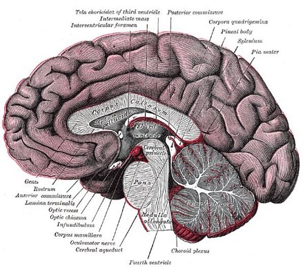
Brain Ventricle Anatomy Diagram Image Radiopaedia Org

Brain Ventricle Anatomy Diagram Image Radiopaedia Org Brain. anatomy by mohamed saber brain. cysts, ventricle, csf by mohamed saber anatomy review brain by craig hacking neuro by rene roberts radiopaedia 2024 virtual conference: cns ventricular lesions by derek smith neuroanatomy and pathology by fraser merchant; unlisted playlists. this case is used in 50 unlisted playlists. Brain ventricle anatomy diagram. case contributed by matt skalski skalski m, brain ventricle anatomy diagram. case study, radiopaedia.org (accessed on 24 nov 2023.

Lateral Ventricle Radiology Reference Article Radiopaedia Org Advertisement: radiopaedia is free thanks to our supporters and advertisers. brain ventricle anatomy diagram. diagram. contributed by matt skalski on june 25, 2015. Ct brain normal ventricles. page author: dr graham lloyd jones ba mbbs mrcp frcr consultant radiologist salisbury nhs foundation trust uk (read bio) ct brain images ct appearances of the cerebral ventricles including the lateral ventricles, third ventricle, fourth ventricle, basal cisterns and cisterna magna. The computer based tutorial, "the cerebral ventricles," enables the user to review the anatomy, imaging, and common pathologic conditions of the human cerebral ventricular system. the program runs on a workstation that includes a laser videodisk player and a videodisk with 21,000 still images plus motion sequences. by using a mouse to select specific portions of the anatomic diagram depicting. It is a narrow slit that is bordered laterally by the medial nuclei of each thalamus, the hypothalamus and interrupted anteriorly by the interthalamic adhesion. the roof of the cavity is formed anteriorly by the fornix and posteriorly by the splenium of the corpus callosum. third ventricle. ventriculus tertius. 1 5.

Ventricular System Radiology Reference Article Radiopaedia Org The computer based tutorial, "the cerebral ventricles," enables the user to review the anatomy, imaging, and common pathologic conditions of the human cerebral ventricular system. the program runs on a workstation that includes a laser videodisk player and a videodisk with 21,000 still images plus motion sequences. by using a mouse to select specific portions of the anatomic diagram depicting. It is a narrow slit that is bordered laterally by the medial nuclei of each thalamus, the hypothalamus and interrupted anteriorly by the interthalamic adhesion. the roof of the cavity is formed anteriorly by the fornix and posteriorly by the splenium of the corpus callosum. third ventricle. ventriculus tertius. 1 5. Aside from cerebrospinal fluid, your brain ventricles are hollow. their sole function is to produce and secrete cerebrospinal fluid to protect and maintain your central nervous system. csf is constantly bathing the brain and spinal column, clearing out toxins and waste products released by nerve cells. This interactive brain model is powered by the wellcome trust and developed by matt wimsatt and jack simpson; reviewed by john morrison, patrick hof, and edward lein. structure descriptions were written by levi gadye and alexis wnuk and jane roskams .

Comments are closed.