Bones And Joints Of The Foot And Ankle Overview Footeducation

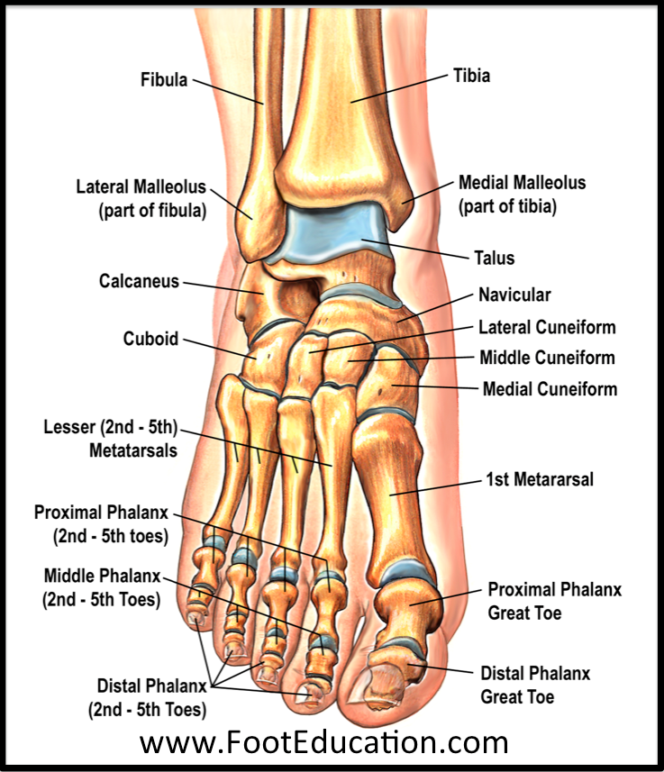
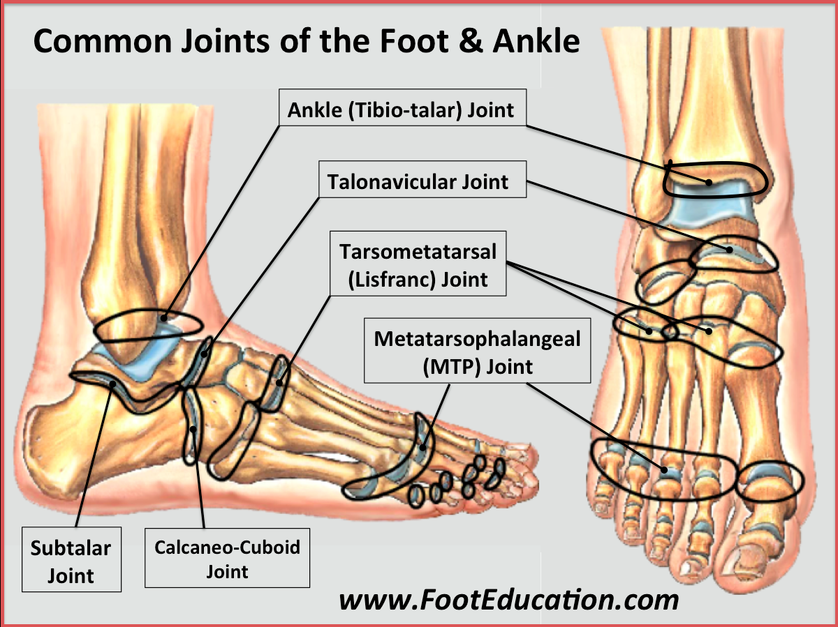
Bones And Joints Of The Foot And Ankle Overview Footeducation The interosseous membrane thickens in the lower part of the leg in order to make the ankle more stable. the tibia and fibula form the ankle joint where they connect with the talus. these two bones form a sort of dish which the talus fits into. this dish is known as the mortise of the ankle joint. Learn more. visual anatomy tool to see foot and ankle joints. footeducation is committed to helping educate patients about foot and ankle conditions by providing high quality, accurate, and easy to understand information.

Bones And Joints Of The Foot And Ankle Overview Footeducation Foot and ankle anatomy. use our anatomy tools to learn about bones, joints, ligaments, and muscles of the foot and ankle. footeducation is committed to helping educate patients about foot and ankle conditions by providing high quality, accurate, and easy to understand information. Ankle anatomy. the ankle joint, also known as the talocrural joint, allows dorsiflexion and plantar flexion of the foot. it is made up of three joints: upper ankle joint (tibiotarsal), talocalcaneonavicular, and subtalar joints. the last two together are called the lower ankle joint. Foot and ankle anatomy consists of 33 bones, 26 joints and over a hundred muscles, ligaments and tendons. this complex network of structures fit and work together to bear weight, allow movement and provide a stable base for us to stand and move on. the foot needs to be strong and stable to support us, yet flexible to allow all sorts of complex. Introduction. a solid understanding of anatomy is essential to effectively diagnose and treat patients with foot and ankle problems. anatomy is a road map. most structures in the foot are fairly superficial and can be easily palpated. anatomical structures (tendons, bones, joints, etc) tend to hurt exactly where they are injured or inflamed.

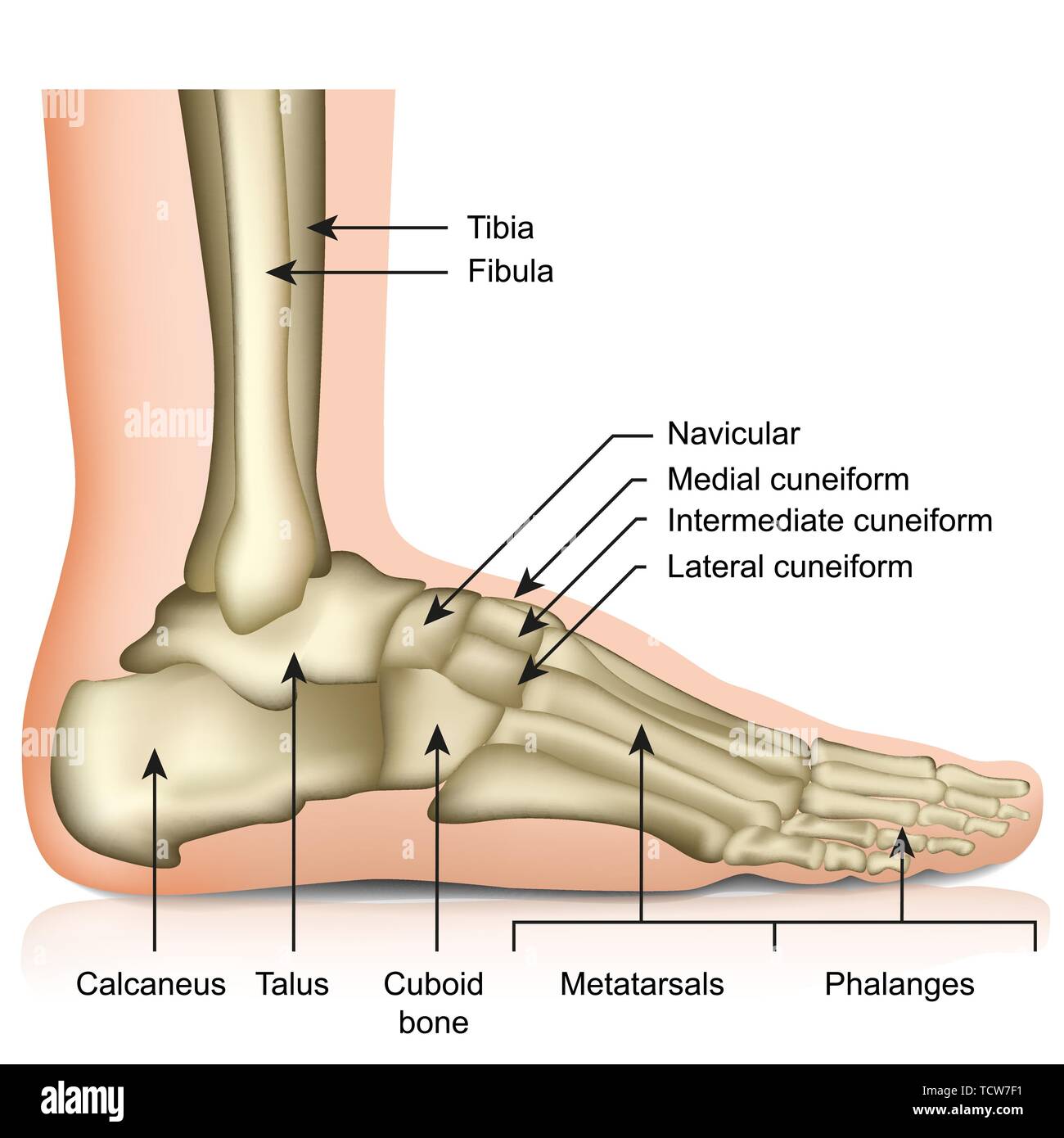
Bones Of The Foot And Ankle Joint Medical Vector Illustration Isolated Foot and ankle anatomy consists of 33 bones, 26 joints and over a hundred muscles, ligaments and tendons. this complex network of structures fit and work together to bear weight, allow movement and provide a stable base for us to stand and move on. the foot needs to be strong and stable to support us, yet flexible to allow all sorts of complex. Introduction. a solid understanding of anatomy is essential to effectively diagnose and treat patients with foot and ankle problems. anatomy is a road map. most structures in the foot are fairly superficial and can be easily palpated. anatomical structures (tendons, bones, joints, etc) tend to hurt exactly where they are injured or inflamed. The foot and ankle form a complex system which consists of 28 bones, 33 joints, 112 ligaments, controlled by 13 extrinsic and 21 intrinsic muscles. the foot is subdivided into the rearfoot, midfoot, and forefoot. it functions as a rigid structure for weight bearing and it can also function as a flexible structure to conform to uneven terrain. In many ways, the structure of the ankle and foot resembles a functional three dimensional puzzle that can be modified, when necessary, to promote either mobility or stability. this chapter provides an overview of the muscles and joints that make up this three dimensional structure.

Comments are closed.