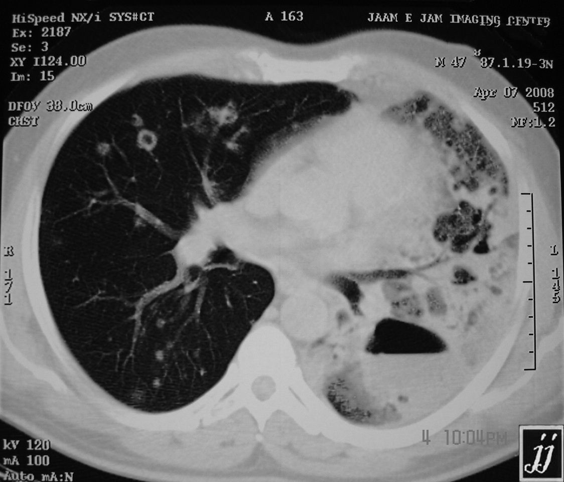
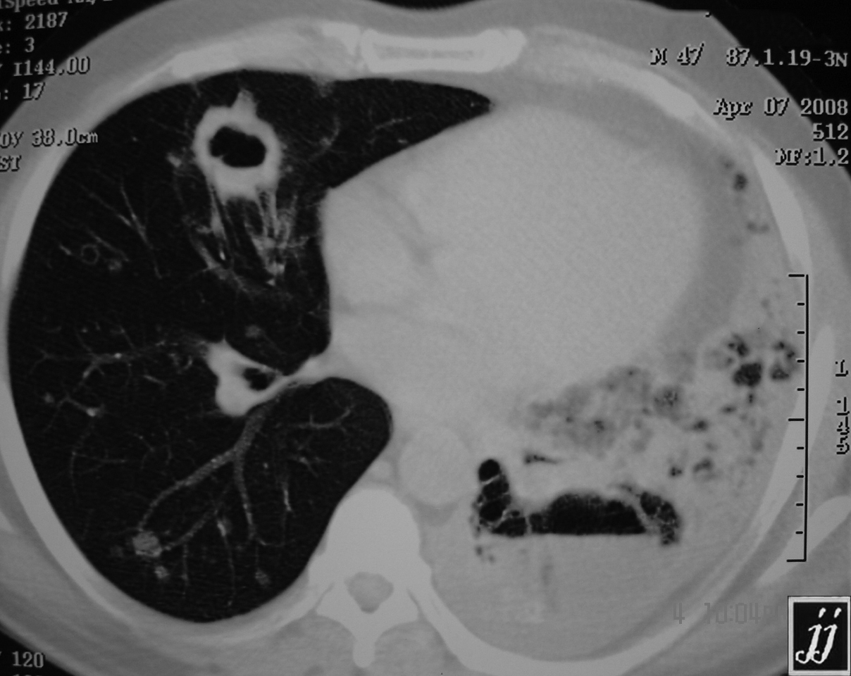
Bilateral Lung Metastasis Radiopaedia

Bilateral Lung Metastasis Radiopaedia Pulmonary metastases are usually asymptomatic or can present with hemoptysis, dyspnea, and pneumothorax. symptoms more commonly arise from the primary tumor, extrapulmonary metastates or systemic effects 5. pulmonary thrombotic microangiopathy is an exception, causing hypoxemia, pulmonary hypertension, cor pulmonale, rapid decline and death 10. Citation, doi, disclosures and article data. cannonball metastases refer to multiple large, well circumscribed, round pulmonary metastases that appear not unsurprisingly like cannonballs. the french terms " envolée de ballons " and " lâcher de ballons ", which translate to "balloons release", are also used to describe this same appearance.

Bilateral Lung Metastasis Radiopaedia Most pulmonary metastases occurring as single or multiple nodules are asymptomatic. when present, symptoms are nonspecific and include cough, hemoptysis, and shortness of breath. the most common clinical manifestation of lymphatic spread of tumor is dyspnea. the dyspnea is typically insidious in onset but tends to progress rapidly. Lung is a common site of metastases; usually multiple, bilateral, sharply outlined, rapidly growing, more pleomorphic and necrotic than lung primaries. may appear as multiple discrete nodules in periphery of lung or as lymphangitic carcinomatosis (peribronchial and perivascular patterns via lymphatics) rarely appear as intralymphatic. The clinical importance of interstitial lung abnormality (ila) is increasingly recognized. in july 2020, the fleischner society published a position paper about ila. the purposes of this article are to summarize the definition, existing evidence, clinical management, and unresolved issues for ila from a radiologic standpoint and to provide a practical guide for radiologists. ila is a common. Typical radiologic findings of a pulmonary metastasis include multiple round variable sized nodules and diffuse thickening of interstitium. in daily practice, however, atypical radiologic features of metastases are often encountered that make distinction of metastases from other nonmalignant pulmonary diseases difficult. a detailed knowledge of the atypical radiologic features of a pulmonary.
A Chest X Ray Pa View Bilateral Lung Cannon Ball Metastasis With The clinical importance of interstitial lung abnormality (ila) is increasingly recognized. in july 2020, the fleischner society published a position paper about ila. the purposes of this article are to summarize the definition, existing evidence, clinical management, and unresolved issues for ila from a radiologic standpoint and to provide a practical guide for radiologists. ila is a common. Typical radiologic findings of a pulmonary metastasis include multiple round variable sized nodules and diffuse thickening of interstitium. in daily practice, however, atypical radiologic features of metastases are often encountered that make distinction of metastases from other nonmalignant pulmonary diseases difficult. a detailed knowledge of the atypical radiologic features of a pulmonary. Cavitation is thought to occur in around 4% of lung metastases 2. pathology. cavitary pulmonary metastases can come from the following primaries: squamous cell carcinoma (70%): lung or head and neck primary 1,4,6. adenocarcinoma: gastrointestinal tract, breast 4,6. sarcoma 4. transitional cell carcinoma of bladder 3. cervical cancer 7. Radiography. the most common radiographic pattern of pulmonary metastasis is the presence of multiple nodules, ranging in size from 3 mm to 15 cm or more. the nodules are more common in the lung bases (because of higher blood flow than upper lobes) and in the outer third of the lungs in the subpleural region.

Pulmonary Metastases Radiology Reference Article Radiopaedia Org Cavitation is thought to occur in around 4% of lung metastases 2. pathology. cavitary pulmonary metastases can come from the following primaries: squamous cell carcinoma (70%): lung or head and neck primary 1,4,6. adenocarcinoma: gastrointestinal tract, breast 4,6. sarcoma 4. transitional cell carcinoma of bladder 3. cervical cancer 7. Radiography. the most common radiographic pattern of pulmonary metastasis is the presence of multiple nodules, ranging in size from 3 mm to 15 cm or more. the nodules are more common in the lung bases (because of higher blood flow than upper lobes) and in the outer third of the lungs in the subpleural region.

Comments are closed.