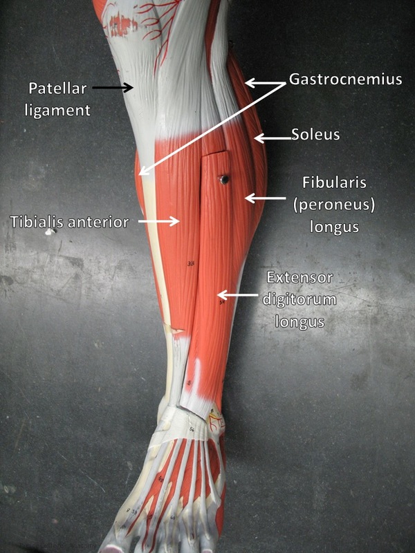
Anterior Lower Leg Muscles Model

Anterior Leg Muscles Anatomy 1 2. synonyms: none. this muscle is the most posterior and lateral of all the muscles of the anterior leg. its origin is lateral and above the origin of the extensor digitorum muscle, precisely in the proximal half of the medial surface of the fibula and lateral condyle and shaft of the tibia. the muscle fibers descend through the anterior. Leg muscles (musculi cruris) anatomically, the leg is defined as the region of the lower limb below the knee. it consists of a posterior, anterior and lateral compartment. in accordance, the muscles of the leg are organized into three groups: anterior (dorsiflexor) group, which contains the tibialis anterior, extensor digitorum longus.

6 Muscles Of The Lower Leg Simplemed Learning Medicine Simplified Explore the anatomy of leg muscles, including anterior and lateral regions, with teachmeanatomy's comprehensive guide. The muscles in the anterior compartment of the leg are a group of four muscles that act to dorsiflex and invert the foot. these muscles are collectively innervated by the deep fibular nerve (l4 s2). the arterial supply is through the anterior tibial artery. in this article, we shall look at the anatomy of the anterior leg muscles – their. Shin muscles, such as the tibialis anterior and extensor digitorum longus, dorsiflex the foot and extend the toes. the muscles of the calf also work subtly to stabilize the ankle joint and foot and to maintain the body's balance. explore the anatomy and function of the leg and foot muscles with innerbody's interactive 3d model. The sartorius is the longest muscle in the body. it is long and thin, running across the thigh in a inferomedial direction. the sartorius is positioned more superficially than the other muscles in the leg. attachments: originates from the anterior superior iliac spine, and attaches to the superior, medial surface of the tibia.

Bio201 Leg Muscles Muscle Anatomy Medical Anatomy Leg Muscles Anatomy Shin muscles, such as the tibialis anterior and extensor digitorum longus, dorsiflex the foot and extend the toes. the muscles of the calf also work subtly to stabilize the ankle joint and foot and to maintain the body's balance. explore the anatomy and function of the leg and foot muscles with innerbody's interactive 3d model. The sartorius is the longest muscle in the body. it is long and thin, running across the thigh in a inferomedial direction. the sartorius is positioned more superficially than the other muscles in the leg. attachments: originates from the anterior superior iliac spine, and attaches to the superior, medial surface of the tibia. The muscles of the lower leg, called simply the leg by anatomists, largely move the foot and toes. the major muscles of the lower leg, other than the gastrocnemius which is cut away, are shown in figure 9.12. the gastrocnemius muscle has two large bellies, called the medial head and the lateral head, and inserts into the calcaneus bone of the. Here’s a leg muscles diagram to give you an overview: as the name suggests, the anterior leg muscles are located along the anterior aspect of the leg. there are four muscles in this compartment: tibialis anterior, extensor hallucis longus, extensor digitorum longus, and fibularis tertius. they receive their innervation via the deep fibular nerve.

Comments are closed.