Anatomy Of The Upper Back Muscles

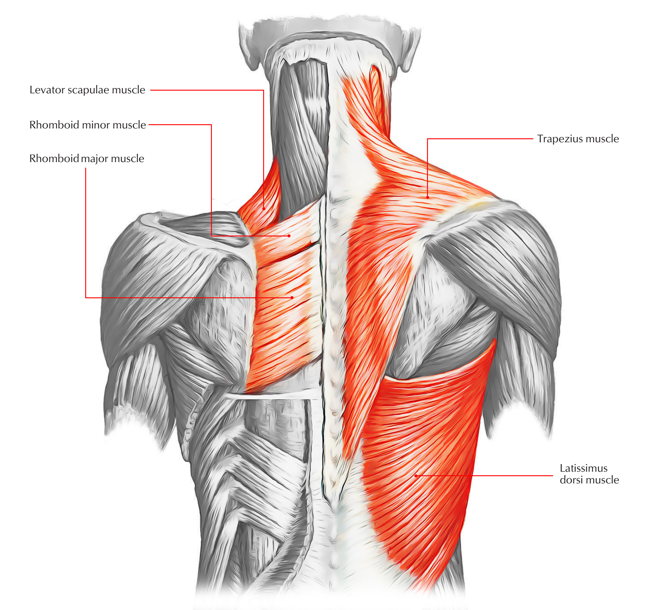
Anatomy Of The Upper Back Muscles Your lat muscles are the largest muscles in the upper half of your body. they start below your shoulder blades and extend to your spine in your lower back. levator scapulae: these are smaller muscles that start at the side of your neck and extend to your shoulder blades. rhomboids: the rhomboid muscles connect your shoulder blades to your spine. The superficial back muscles are the muscles found just under the skin. within this group of back muscles you will find the latissimus dorsi, the trapezius, levator scapulae and the rhomboids. these muscles are able to move the upper limb as they originate at the vertebral column and insert onto either the clavicle, scapula or humerus. whilst.

Upper Back Anatomy Fitness Workouts Exercises The back muscles are divided into two large groups: the extrinsic (superficial) back muscles, which lie most superficially on the back. these muscles are also called immigrant muscles, since they actually represent muscles of the upper limb that have migrated to the back during fetal development. these muscles are divided into superficial and. This is the anatomy of your back muscles, plus how to train them. your latissimus dorsi, or lats, are the largest individual muscles in your upper back. they run down the sides of your torso. Back anatomy. the back is the body region between the neck and the gluteal regions. it comprises the vertebral column (spine) and two compartments of back muscles; extrinsic and intrinsic. the back functions are many, such as to house and protect the spinal cord, hold the body and head upright, and adjust the movements of the upper and lower limbs. Explore the anatomy and function of the chest and upper back muscles with innerbody's interactive 3d model. the muscles of the chest and upper back occupy the thoracic region of the body inferior to the neck and superior to the abdominal region and include the muscles of the shoulders. these important muscles control many motions that involve.

Anatomy Of Back Muscles Diagram Quizlet Back anatomy. the back is the body region between the neck and the gluteal regions. it comprises the vertebral column (spine) and two compartments of back muscles; extrinsic and intrinsic. the back functions are many, such as to house and protect the spinal cord, hold the body and head upright, and adjust the movements of the upper and lower limbs. Explore the anatomy and function of the chest and upper back muscles with innerbody's interactive 3d model. the muscles of the chest and upper back occupy the thoracic region of the body inferior to the neck and superior to the abdominal region and include the muscles of the shoulders. these important muscles control many motions that involve. Summary. the back consists of the spine, spinal cord, muscles, ligaments, and nerves. these structures work together to support the body, enable a range of movements, and send messages from the. Treatment. your back muscles support your spine, attach your pelvis and shoulders to your trunk, and provide mobility and stability to your trunk and spine. the five major muscles of the back are the trapezius, latissimus dorsi, the rhomboids, the erector spinae, and the levator scapulae. back muscles are grouped into three categories.

The Superficial Back Muscles Attachments Actions Teachmeanatomy Summary. the back consists of the spine, spinal cord, muscles, ligaments, and nerves. these structures work together to support the body, enable a range of movements, and send messages from the. Treatment. your back muscles support your spine, attach your pelvis and shoulders to your trunk, and provide mobility and stability to your trunk and spine. the five major muscles of the back are the trapezius, latissimus dorsi, the rhomboids, the erector spinae, and the levator scapulae. back muscles are grouped into three categories.

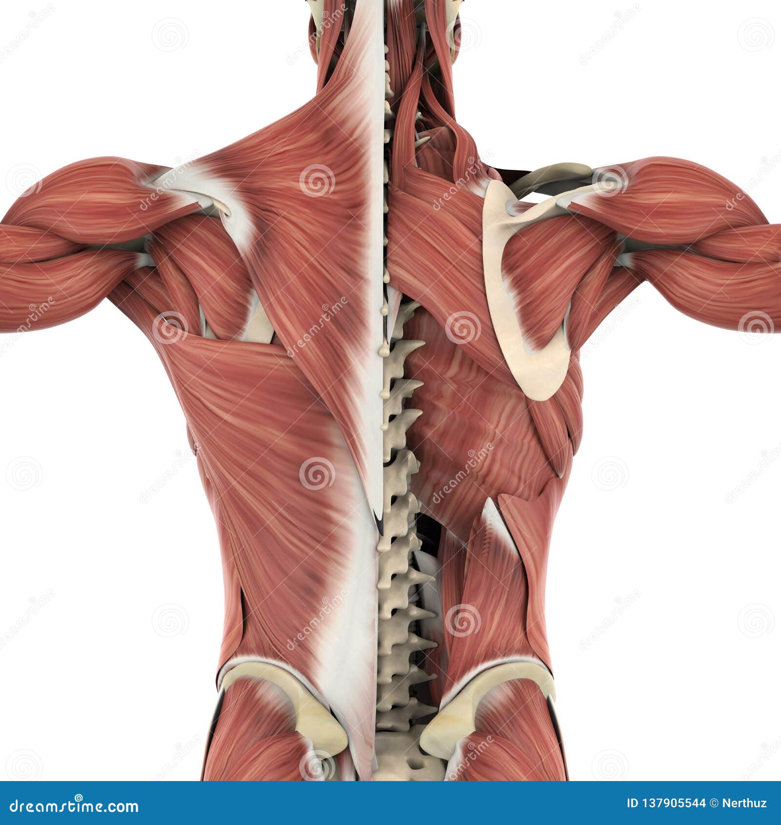
Muscles Of The Back Anatomy Stock Illustration Illustration Of

Comments are closed.