Anatomy Of The Collateral Ligaments Of The Knee Joint Youtube

Anatomy Of The Collateral Ligaments Of The Knee Joint Youtube The knee is the largest joint in the human body. it is a compound joint. not only is it where the femur, or thigh bone, meets the tibia, or shin bone – at th. In this video prof bellemans explains which 5 ligaments stabilize the knee, how they work, and how they may get damaged, stretched or tear.00:00 anatomy of t.

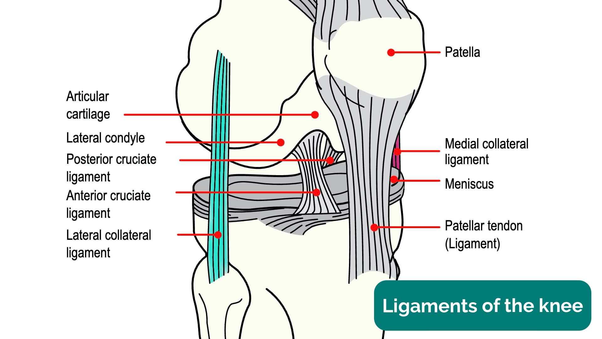
Anatomy Of The Knee Joint Youtube Detailed discussion on anatomy and biomechanics of medial collateral and lateral collateral ligaments of knee complex.medial collateral ligament origins from. The medial collateral ligament (mcl) is on the inner side of your knee. it attaches the thigh bone (femur) to the shin bone (tibia). the lateral collateral ligament (lcl) is on the outer side of your knee. it connects your femur to your calf bone (fibula). the collateral ligaments prevent the knee from moving side to side too much. cruciate. The tibial collateral ligament is the strong, flat ligament of the medial aspect of the knee joint. the tibial collateral ligament, in addition to its fibular counterpart, acts to secure the knee joint and prevent excessive sideways movement by restricting external and internal rotation of the extended knee. The knee joint is the biggest joint in your body. it connects your thigh bone (femur) to your shin bone (tibia). it helps you stand, move and keep your balance. your knees also contain cartilage, like your meniscus, and ligaments, including your lcl, mcl, acl and pcl. find a primary care provider. schedule an appointment.

Knee Joint Anatomy Youtube The tibial collateral ligament is the strong, flat ligament of the medial aspect of the knee joint. the tibial collateral ligament, in addition to its fibular counterpart, acts to secure the knee joint and prevent excessive sideways movement by restricting external and internal rotation of the extended knee. The knee joint is the biggest joint in your body. it connects your thigh bone (femur) to your shin bone (tibia). it helps you stand, move and keep your balance. your knees also contain cartilage, like your meniscus, and ligaments, including your lcl, mcl, acl and pcl. find a primary care provider. schedule an appointment. It’s sometimes also called the lateral collateral ligament, or the fcl, and you’ll notice that abbreviating is a running theme with ligaments of the knee joint. the fibular collateral ligament is a strong cord like ligament stretching from the lateral epicondyle of the femur to the fibular head, and it’s separated from the joint capsule. The anterior cruciate ligament (acl) is one of the most frequently injured ligaments in the knee. ligaments are strong, non elastic fibers that connect bones. the acl, which runs inside the knee, connects the thighbone (femur) to the shinbone (tibia), providing stability to the knee joint. acl tears often happen to athletes or active.

Knee Joint Anatomy Geeky Medics It’s sometimes also called the lateral collateral ligament, or the fcl, and you’ll notice that abbreviating is a running theme with ligaments of the knee joint. the fibular collateral ligament is a strong cord like ligament stretching from the lateral epicondyle of the femur to the fibular head, and it’s separated from the joint capsule. The anterior cruciate ligament (acl) is one of the most frequently injured ligaments in the knee. ligaments are strong, non elastic fibers that connect bones. the acl, which runs inside the knee, connects the thighbone (femur) to the shinbone (tibia), providing stability to the knee joint. acl tears often happen to athletes or active.

Medial Lateral Collateral Ligaments Anatomy Biomechanics Applied

Comments are closed.