Anatomy And Structure Of The Pectoral Muscles Vector Image

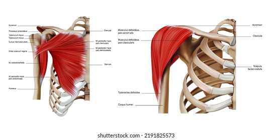
Anatomy And Structure Of The Pectoral Muscles Vector Image Anatomy and structure of the pectoral muscles of the trunk on a white background. vector 3d illustration. download a free preview or high quality adobe illustrator (ai), eps, pdf vectors and high res jpeg and png images. Comprehensive anatomy of the pectoral region including origin, course, and distribution of the major vessels and their branches that supply drain it. muscle innervations from the brachial plexus, together with their sensory distribution to predict or localize the consequences of injury to these nerves along with an explanation on how to test their functional integrity clinically.

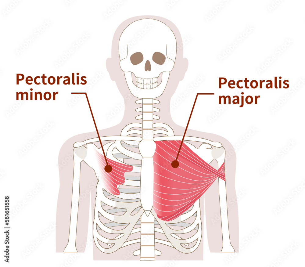
Anatomy Structure Shoulder Pectoral Muscles Trunk Stock Vector Royalty The pectoralis minor lies underneath its larger counterpart muscle, pectoralis major. both muscles form part of the anterior wall of the axilla region. attachments: originates from the 3rd 5th ribs and inserts into the coracoid process of the scapula. function: stabilises the scapula by drawing it anteroinferiorly against the thoracic wall. The pectoralis major has a broad origin, based on which it is divided into three parts: clavicular part, sternocostal part and abdominal part. all three parts converge laterally and insert onto the greater tubercle of humerus. the main function of this chest muscle as a whole is the adduction and internal rotation of the arm in the shoulder joint. The pectoral muscles are the group of skeletal muscles that connect the upper extremities to the anterior and lateral thoracic walls. juxtaposed with the regional fascia, these muscles are responsible for moving the upper extremities in a wide range of motion. these include but are not limited to flexion, adduction, and internal rotation of the humerus, stabilization of the scapula, as well as. The pectoralis major is a muscle of the anterior chest wall. it is a large fan shaped muscle, which is composed of a sternal head and a clavicular head. clavicular head originates from the anterior surface of the medial clavicle. sternocostal head originates from the anterior surface of the sternum, the superior six costal cartilages and the.

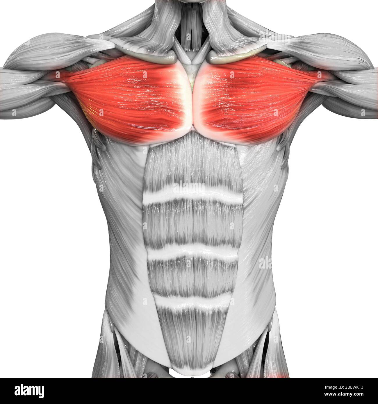
Human Muscular System Parts Pectoral Muscle Anatomy Stock Photo Alamy The pectoral muscles are the group of skeletal muscles that connect the upper extremities to the anterior and lateral thoracic walls. juxtaposed with the regional fascia, these muscles are responsible for moving the upper extremities in a wide range of motion. these include but are not limited to flexion, adduction, and internal rotation of the humerus, stabilization of the scapula, as well as. The pectoralis major is a muscle of the anterior chest wall. it is a large fan shaped muscle, which is composed of a sternal head and a clavicular head. clavicular head originates from the anterior surface of the medial clavicle. sternocostal head originates from the anterior surface of the sternum, the superior six costal cartilages and the. The pectoralis major is a thick, fan shaped muscle, situated at the upper and forepart of the chest. it arises from the anterior surface of the sternal half of the clavicle; from half the breadth of the anterior surface of the sternum, as low down as the attachment of the cartilage of the sixth or seventh rib; from the cartilages of all the. The pectoralis major is the superior most and largest muscle of the anterior chest wall. it is a thick, fan shaped muscle that lies underneath the breast tissue and forms the anterior wall of the axilla. its origin lies anterior surface of the medial half of the clavicle, the anterior surface of the sternum, the first 7 costal cartilages, the sternal end of the sixth rib, and the aponeurosis.

Illustration Of The Anatomy Of The Pectoralis Major And Minor Muscle The pectoralis major is a thick, fan shaped muscle, situated at the upper and forepart of the chest. it arises from the anterior surface of the sternal half of the clavicle; from half the breadth of the anterior surface of the sternum, as low down as the attachment of the cartilage of the sixth or seventh rib; from the cartilages of all the. The pectoralis major is the superior most and largest muscle of the anterior chest wall. it is a thick, fan shaped muscle that lies underneath the breast tissue and forms the anterior wall of the axilla. its origin lies anterior surface of the medial half of the clavicle, the anterior surface of the sternum, the first 7 costal cartilages, the sternal end of the sixth rib, and the aponeurosis.

Comments are closed.